
| Download the amazing global Makindo app: Android | Apple | |
|---|---|
| MEDICAL DISCLAIMER: Educational use only. Not for diagnosis or management. See below for full disclaimer. |
Acute Abdominal Pain - Adults
Related Subjects:Acute Cholecystitis |Acute Appendicitis |Chronic Peritonitis |Abdominal Aortic Aneurysm |Ectopic Pregnancy |Acute Cholangitis |Acute Abdominal Pain/Peritonitis |Assessing Abdominal Pain |Penetrating Abdominal Trauma
🫀 Peritonitis is a surgical emergency caused by inflammation of the peritoneum, often due to perforated abdominal organs (e.g., appendicitis, diverticulitis, peptic ulcer, ruptured AAA). ⚠️ Immediate attention to ABC (airway, breathing, circulation) is critical. 🚨 In elderly, immunocompromised, or steroid-treated patients, signs may be subtle or absent. ⏱️ Early diagnosis and surgical intervention reduce morbidity and mortality.
| 🩺 Initial Management Summary |
|---|
|
ℹ️ About
- 🌊 Generalised Peritonitis: Due to perforated viscus → widespread inflammation.
- 🦠 Acute Suppurative: Pus formation from bacterial infection → classic acute abdomen presentation.
🧪 Aetiology
- 🩸 Inflammation of Peritoneum: Triggered by infection, chemical irritation, or trauma.
- ⚠️ Causes: Leakage of gastric contents, bile, blood, or bacteria into peritoneal cavity.
- 🧪 Aseptic vs. Septic: Sterile irritants (bile, acid, pancreatic juice) may become secondarily infected.
📊 Classifications
- ⏱️ Acute vs. Chronic: Acute = emergency; Chronic (e.g., TB) = slow onset.
- 🔍 Primary vs. Secondary:
- Primary = haematogenous spread (e.g., SBP in cirrhosis).
- Secondary = intra-abdominal source (perforated organ, postop leak).
- 🧫 Septic vs. Aseptic: Bacterial vs. sterile causes (e.g., pancreatitis).
🚨 Causes of Acute Peritonitis
- 🍽️ GI perforations: Appendicitis rupture, diverticulitis, perforated ulcers.
- 💚 Biliary/hepatic: Gallbladder rupture, bile leak.
- 🤕 Trauma: Penetrating injuries, anastomotic leaks.
- 👩🍼 Gynaecological: Ectopic pregnancy rupture, pelvic infection.
- 🧴 Spontaneous Bacterial Peritonitis: In cirrhotic ascites.
- 🌍 Infectious: TB, typhoid, dialysis-related peritonitis.
🧫 Microbiology
- Gram−: E. coli, Klebsiella, Bacteroides fragilis
- Gram+: Streptococcus, Enterococcus
- Fungal: Candida (esp. immunosuppressed)
- Chronic: Mycobacterium tuberculosis
🩺 Clinical Features
- 😖 Severe sudden abdominal pain ± shoulder tip pain (diaphragmatic irritation)
- 🤮 Nausea, vomiting, distension
- 🛡️ Rigid “board-like” abdomen, guarding, rebound
- 📈 Fever, tachycardia, hypotension
- 💥 Septic shock in severe cases
🔎 Investigations
- 🧪 FBC (↑WCC), CRP, U&E, LFTs, lactate, amylase, blood cultures
- ❤️ ECG (exclude MI, look for AF/embolus)
- 👩🍼 β-hCG in women of childbearing age
- 🩻 CXR (free air under diaphragm)
- 🖥️ CT (gold standard for diagnosis and localisation)
- 🩺 USS (abscess/biliary pathology)
⚠️ Complications
- 🦠 Sepsis & septic shock
- 🔥 SIRS → ARDS, AKI
- ⛔ Paralytic ileus
- 💔 Multiorgan failure, death
⚡ Acute Abdominal Pain in Adults - Causes, Features, Investigations & Management
Causes of acute abdominal pain can be grouped by system. This table summarises key conditions with their classic presentation, investigations, and first-line management.
| Cause | Clinical Features | Investigations | Management |
|---|---|---|---|
| Appendicitis 🟠 | Periumbilical → RLQ pain, nausea, fever, rebound tenderness | FBC, CRP, urinalysis, US/CT abdomen | IV fluids, antibiotics, appendicectomy |
| Diverticulitis 🟡 | LLQ pain, fever, altered bowel habit | FBC, CRP, CT abdomen with contrast | IV antibiotics, fluids, surgery if perforated/abscess |
| Perforated Peptic Ulcer 🔴 | Sudden severe epigastric pain, peritonitis, rigid abdomen | Upright CXR (free air), CT abdomen | Resuscitation, IV antibiotics, emergency laparotomy |
| Small Bowel Obstruction 🚫 | Colicky central pain, vomiting, distension, constipation | AXR (dilated loops, air-fluid levels), CT abdomen | NG tube decompression, IV fluids, surgery if strangulated |
| Mesenteric Ischaemia ⚡ | Severe pain “out of proportion”, AF history, metabolic acidosis | Lactate, CT angiography | IV fluids, antibiotics, urgent revascularisation/resection |
| Cholecystitis 🟢 | RUQ pain, fever, Murphy’s sign | US abdomen (stones, wall thickening), LFTs | IV antibiotics, fluids, laparoscopic cholecystectomy |
| Cholangitis 🟢 | Charcot’s triad: RUQ pain, fever, jaundice | LFTs (↑ ALP, GGT, bilirubin), US/MRCP | IV antibiotics, ERCP for stone removal |
| Pancreatitis 🔥 | Severe epigastric pain radiating to back, nausea, vomiting | Serum amylase/lipase ↑, CT abdomen for severity | NBM, IV fluids, analgesia, treat cause (e.g., gallstones, alcohol) |
| Renal Colic 💎 | Severe loin-to-groin pain, haematuria, restlessness | Urinalysis, CT KUB (non-contrast) | Analgesia (NSAIDs), fluids, urology referral if obstructed |
| Ruptured AAA 💥 | Sudden severe abdominal/back pain, shock, pulsatile mass | Bedside US (rapid), CT if stable | ABC resuscitation, emergency vascular surgery |
✅ Key Clinical Advice
- Always assess and stabilise with ABCDE before focusing on diagnosis.
- In unstable patients → urgent surgical review, do not delay for imaging.
- Consider broad differentials; elderly and immunosuppressed may have atypical presentations.
💊 Management
- 🔄 Resuscitation: ABCs, O₂, IV fluids, catheter for urine output.
- 💉 Antibiotics: Broad-spectrum IV (e.g., Tazocin ± metronidazole).
- 💊 Analgesia: IV morphine (2.5–5 mg), antiemetics.
- 🥛 NBM: Prepare for surgery.
- 🧑⚕️ Urgent surgical referral: Early laparotomy/laparoscopy.
🔪 Surgical Management
- Indicated for perforated viscus, bowel ischaemia, or generalised peritonitis.
- Procedures:
- Exploratory laparotomy/laparoscopy
- Resection of necrotic bowel, repair perforations
- Peritoneal lavage, drain abscesses
- Continue IV antibiotics post-op.
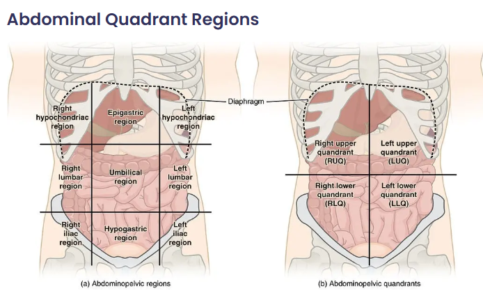
⚡ Acute Abdominal Pain in Adults
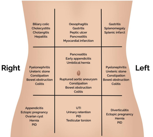
Acute abdominal pain is a common emergency presentation. Causes vary by anatomical location. Careful history, examination, and prompt investigations guide diagnosis and management.
| Region & Causes | Clinical Features | Investigations | Management |
|---|---|---|---|
| Right Upper Quadrant (RUQ) 🟢
- Cholecystitis - Cholangitis - Hepatitis - Peptic ulcer perforation |
- RUQ pain ± radiation to shoulder
- Murphy’s sign (cholecystitis) - Jaundice, fever (Charcot’s triad → cholangitis) |
- LFTs (↑ ALP, GGT, bilirubin)
- Abdominal US (gallstones, biliary dilatation) - MRCP/ERCP if obstructive |
- Analgesia, IV fluids
- IV antibiotics (if infection) - Cholecystectomy (definitive for cholecystitis) - ERCP for obstructive stones |
| Left Upper Quadrant (LUQ) 🔵
- Splenic infarct/rupture - Gastritis/peptic ulcer - Pancreatitis |
- LUQ pain ± referred to left shoulder (Kehr’s sign)
- History of trauma (rupture) - Epigastric pain radiating to back (pancreatitis) |
- FBC, amylase/lipase (↑ in pancreatitis)
- CT abdomen (trauma, pancreatitis severity) - Endoscopy (if suspected ulcer/gastritis) |
- Supportive: fluids, analgesia, NBM
- Pancreatitis → IV fluids, monitor severity - Splenic rupture → urgent surgery if unstable |
| Right Lower Quadrant (RLQ) 🟠
- Appendicitis - Mesenteric adenitis - Crohn’s flare - Renal colic |
- Periumbilical → RLQ pain
- Nausea, anorexia, fever - Rovsing’s sign, rebound tenderness |
- FBC (↑ WCC, CRP)
- Urinalysis (exclude UTI/renal colic) - US or CT (appendicitis, renal stones) |
- Appendicectomy (surgical)
- IV antibiotics - Fluids and analgesia |
| Left Lower Quadrant (LLQ) 🟡
- Diverticulitis - Sigmoid volvulus - Colon cancer - Renal colic |
- LLQ pain ± fever
- Altered bowel habit - Abdominal distension (volvulus) |
- FBC, CRP
- CT abdomen/pelvis with contrast (gold standard for diverticulitis) - AXR (coffee-bean sign in volvulus) |
- IV antibiotics, fluids (diverticulitis)
- Endoscopic decompression or surgery (volvulus) - Refer colorectal if malignancy suspected |
| Epigastric/Generalised 🔴
- Perforated peptic ulcer - Pancreatitis - Small bowel obstruction - Mesenteric ischaemia |
- Sudden severe “knife-like” pain (perforation)
- Vomiting, distension (obstruction) - Severe, disproportionate pain (ischaemia) |
- FBC, U&E, lactate (↑ in ischaemia)
- Amylase/lipase (pancreatitis) - AXR/CT (free air, obstruction, ischaemia) |
- ABC resuscitation
- IV fluids, NG tube decompression - Broad-spectrum antibiotics - Emergency laparotomy if perforation or ischaemia |
✅ Key Points
- Always assess ABCDE and resuscitate if unstable.
- Localise pain anatomically to guide differential diagnosis.
- Bloods + Urinalysis + Imaging form the core of investigation.
- Early surgical review is essential for suspected perforation, obstruction, ischaemia, or appendicitis.
📚 References
- Peritonitis: Pathophysiology & Management
- UpToDate: Acute Peritonitis
- Medscape: Peritonitis Overview
Case 1 – Perforated duodenal ulcer
A 48-year-old man with NSAID use develops sudden, severe epigastric pain radiating to the shoulder, rigid abdomen, and absent bowel sounds; vitals show tachycardia, low-grade fever, and mild hypotension. Erect CXR shows free subdiaphragmatic air. Manage with ABCDE, IV fluids, broad-spectrum antibiotics, PPI, NG tube, and urgent CT abdomen; refer for emergency surgery (laparoscopic Graham patch vs definitive ulcer surgery) and test/treat H. pylori after recovery.
Case 2 – Ruptured abdominal aortic aneurysm (AAA)
A 72-year-old man with smoking and hypertension presents with sudden tearing back/abdominal pain, hypotension, and a pulsatile abdominal mass. Avoid excessive fluids (permissive hypotension), give O₂, activate massive haemorrhage protocol, crossmatch, and call vascular surgery for immediate EVAR/open repair. POCUS/bedside ultrasound confirms large infrarenal AAA; do not delay for CT if unstable.
Case 3 – Ruptured ectopic pregnancy
A 30-year-old with 7 weeks’ amenorrhoea and vaginal spotting presents with worsening lower abdominal pain, dizziness, and shoulder tip pain; she is tachycardic and hypotensive with abdominal guarding. β-hCG positive; FAST scan shows free fluid. Resuscitate (O₂, IV access, bloods/crossmatch), consult gynae for urgent salpingectomy (or salpingostomy if appropriate), give anti-D if rhesus negative, and manage pain; differentials include ovarian torsion and ruptured corpus luteum.
Case 4 – Acute appendicitis
A 24-year-old develops periumbilical pain migrating to the RIF with anorexia, mild fever, and rebound tenderness; WBC/CRP raised. Ultrasound (slim female) or CT abdomen confirms inflamed, non-compressible appendix. Give IV fluids, analgesia, and broad-spectrum antibiotics; proceed to laparoscopic appendicectomy. Consider differentials (gynae, mesenteric adenitis); beware atypical retrocaecal pain.
Case 5 – Acute mesenteric ischaemia
A 78-year-old with AF has sudden, severe, diffuse abdominal pain out of proportion to scant early signs; lactate rising, metabolic acidosis. Urgent CTA shows SMA embolus. Resuscitate, start IV heparin and broad-spectrum antibiotics, involve vascular/HPB surgery for embolectomy/revascularisation ± bowel resection if non-viable; mortality is high-act fast.
Case 6 – Sigmoid volvulus
An 82-year-old in a care home presents with abdominal distension, pain, constipation, and tympany; X-ray shows the classic coffee-bean sign pointing to the RUQ. If no peritonitis/ischemia, perform flexible sigmoidoscopy with decompression and rectal tube; arrange definitive surgery (e.g., sigmoid colectomy) due to recurrence risk. If peritonitis or perforation, urgent laparotomy.
| The content on this website is provided for educational and informational purposes only to support exam preparation (e.g., MLA, MRCP, USMLE) and learning. This is NOT medical advice, diagnosis, treatment, or professional guidance. It does not replace consultation with a qualified healthcare professional, official guidelines (e.g., NICE, GMC, BNF), or supervised clinical practice. Always verify information with current, authoritative sources. Makindo and its contributors accept no liability for any reliance on this content, including errors, omissions, or any resulting harm, loss, or consequences. By using this site, you agree to these terms. |
|
|
Categories
- About
- Acute Medicine
- Anaesthetics and Critical Care
- Anatomy
- Anatomy and Physiology
- Biochemistry
- Book
- Cardiology
- Collections
- CompSci
- Crib Sheets
- Critical care
- Dental
- Dermatology
- Differentials
- Drugs
- ENT
- Electrocardiogram
- Embryology
- Emergency Medicine
- Endocrinology
- Ethics
- Foundation Doctors
- GCSE
- Gastroenterology
- General Practice
- Genetics
- Geriatric Medicine
- Geriatrics
- Guidelines
- Haematology
- Hepatology
- Immunology
- Infectious Diseases
- Infographic
- Investigations
- Lists
- MRCP
- Mandatory Training
- Medical Students
- Microbiology
- Nephrology
- Neurology
- Neurosurgery
- Nutrition
- OSCE
- Obstetrics Gynaecology
- Oncology
- Ophthalmology
- Oral Medicine and Dentistry
- Orthopaedics
- Paediatrics
- Palliative
- Palliative Care
- Pathology
- Pharmacology
- Physiology
- Procedures
- Psychiatry
- Public Health
- Radiology
- Respiratory
- Resuscitation
- Revision Topics
- Rheumatology
- Statistics and Research
- Stroke
- Surgery
- Toxicology
- Trauma and Orthopaedics
- USMLE
- Urology
- Vascular Surgery