
| Download the amazing global Makindo app: ✅ Means NICE/National Guidelines 2026 compliant Android | Apple | |
|---|---|
| MEDICAL DISCLAIMER: Educational use only. Not for diagnosis or management. See below for full disclaimer. |
ECG - Causes of a Dominant R wave in V1 ❤️
Related Subjects: |ECG Basics |ECG Axis |ECG Analysis |ECG LAD |ECG RAD |ECG Low voltage |ECG Pathological Q waves |ECG ST/T wave changes |ECG LBBB |ECG RBBB |ECG short PR |ECG Heart Block |ECG Asystole and P wave asystole |ECG QRS complex |ECG ST segment |ECG: QT interval |ECG: LVH |ECG RVH |ECG: Bundle branch blocks |ECG Dominant R wave in V1 |ECG Acute Coronary Syndrome |ECG Crib sheets
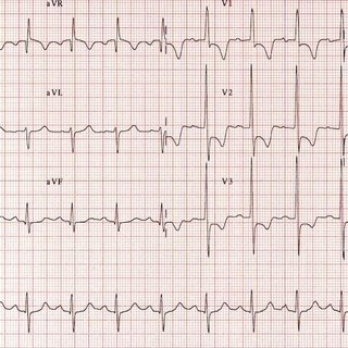
🔎 Causes of a Tall R Wave in V1
- 💪 Right Ventricular Hypertrophy (RVH): Increased RV muscle mass → tall R wave in V1. ⚠️ Seen in pulmonary hypertension (COPD, PE, congenital heart disease).
- ❤️ Posterior Myocardial Infarction: Loss of posterior LV forces produces a reciprocal tall R in V1. ⚠️ Often with ST depression & upright T in V1–V3 → mimics RVH.
- ⚡ Right Bundle Branch Block (RBBB): Delayed RV activation → prominent terminal R in V1. ⚠️ May be isolated or due to structural/ischaemic disease.
- 🔄 Wolff-Parkinson-White (WPW): Pre-excitation via left-sided accessory pathway (Type A) → tall R in V1. ⚠️ Associated with SVT; look for short PR + delta wave.
- 🧬 Hypertrophic Cardiomyopathy (HCM): Asymmetric septal hypertrophy can cause dominant R in V1. ⚠️ Risk of HF & sudden cardiac death; family history key.
- 🙂 Normal Variant: Some healthy young individuals have a tall R in V1. ⚠️ No further workup if asymptomatic and no other ECG abnormalities.
- ➡️ Dextrocardia: Heart on right side of chest → mirror image ECG. ⚠️ Confirm with CXR or echo; look for reversed limb lead polarity.
- 🧠 Neuromuscular Disorders: Duchenne & Myotonic dystrophy can cause ECG changes incl. tall R in V1. ⚠️ Check CK, look for weakness & systemic features.
| The content on this website is provided for educational and informational purposes only to support exam preparation (e.g., MLA, MRCP, USMLE) and learning. This is NOT medical advice, diagnosis, treatment, or professional guidance. It does not replace consultation with a qualified healthcare professional, official guidelines (e.g., NICE, GMC, BNF), or supervised clinical practice. Always verify information with current, authoritative sources. Makindo and its contributors accept no liability for any reliance on this content, including errors, omissions, or any resulting harm, loss, or consequences. By using this site, you agree to these terms. |
|
|
Categories
- About
- Acute Medicine
- Anaesthetics and Critical Care
- Anatomy
- Anatomy and Physiology
- Biochemistry
- Book
- Cardiology
- Collections
- CompSci
- Crib Sheets
- Critical care
- Dental
- Dermatology
- Differentials
- Drugs
- ENT
- Electrocardiogram
- Embryology
- Emergency Medicine
- Endocrinology
- Ethics
- Foundation Doctors
- GCSE
- Gastroenterology
- General Practice
- Genetics
- Geriatric Medicine
- Geriatrics
- Guidelines
- Haematology
- Hepatology
- Immunology
- Infectious Diseases
- Infographic
- Investigations
- Lists
- MRCP
- Mandatory Training
- Medical Students
- Microbiology
- Nephrology
- Neurology
- Neurosurgery
- Nutrition
- OSCE
- Obstetrics Gynaecology
- Oncology
- Ophthalmology
- Oral Medicine and Dentistry
- Orthopaedics
- Paediatrics
- Palliative
- Palliative Care
- Pathology
- Pharmacology
- Physiology
- Procedures
- Psychiatry
- Public Health
- Radiology
- Respiratory
- Resuscitation
- Revision Topics
- Rheumatology
- Statistics and Research
- Stroke
- Surgery
- Toxicology
- Trauma and Orthopaedics
- USMLE
- Urology
- Vascular Surgery