Makindo Medical Notes"One small step for man, one large step for Makindo" |
|
|---|---|
| Download all this content in the Apps now Android App and Apple iPhone/Pad App | |
| MEDICAL DISCLAIMER: The contents are under continuing development and improvements and despite all efforts may contain errors of omission or fact. This is not to be used for the assessment, diagnosis, or management of patients. It should not be regarded as medical advice by healthcare workers or laypeople. It is for educational purposes only. Please adhere to your local protocols. Use the BNF for drug information. If you are unwell please seek urgent healthcare advice. If you do not accept this then please do not use the website. Makindo Ltd. |
ECG - Collection B
-
| About | Anaesthetics and Critical Care | Anatomy | Biochemistry | Cardiology | Clinical Cases | CompSci | Crib | Dermatology | Differentials | Drugs | ENT | Electrocardiogram | Embryology | Emergency Medicine | Endocrinology | Ethics | Foundation Doctors | Gastroenterology | General Information | General Practice | Genetics | Geriatric Medicine | Guidelines | Haematology | Hepatology | Immunology | Infectious Diseases | Infographic | Investigations | Lists | Microbiology | Miscellaneous | Nephrology | Neuroanatomy | Neurology | Nutrition | OSCE | Obstetrics Gynaecology | Oncology | Ophthalmology | Oral Medicine and Dentistry | Paediatrics | Palliative | Pathology | Pharmacology | Physiology | Procedures | Psychiatry | Radiology | Respiratory | Resuscitation | Rheumatology | Statistics and Research | Stroke | Surgery | Toxicology | Trauma and Orthopaedics | Twitter | Urology
Related Subjects: |ECG Basics |ECG Axis |ECG Analysis |ECG LAD |ECG RAD |ECG Low voltage |ECG Pathological Q waves |ECG ST/T wave changes |ECG LBBB |ECG RBBB |ECG short PR |ECG Heart Block |ECG Asystole and P wave asystole |ECG QRS complex |ECG ST segment |ECG: QT interval |ECG: LVH |ECG RVH |ECG: Bundle branch blocks |ECG Dominant R wave in V1 |ECG Acute Coronary Syndrome |ECG Narrow complex tachycardia |ECG Ventricular fibrillation |ECG Regular Broad complex tachycardia |ECG Crib sheets
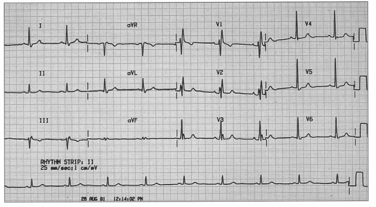
NORMAL
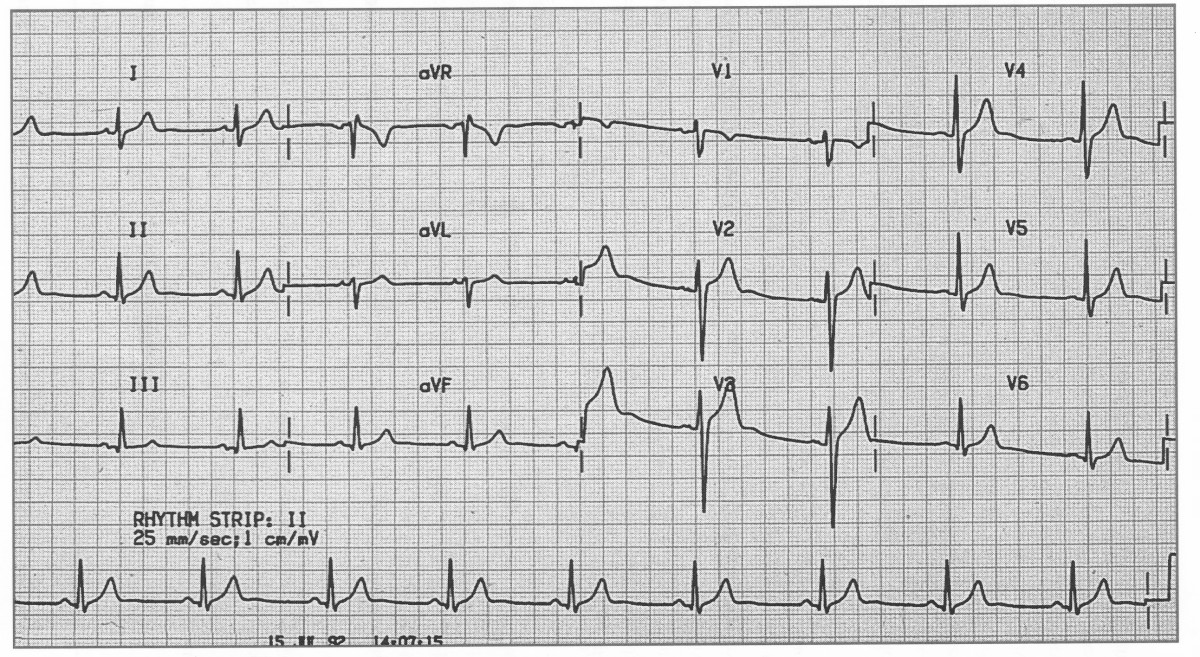
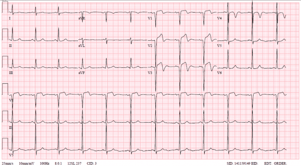
Wellens Syndrome
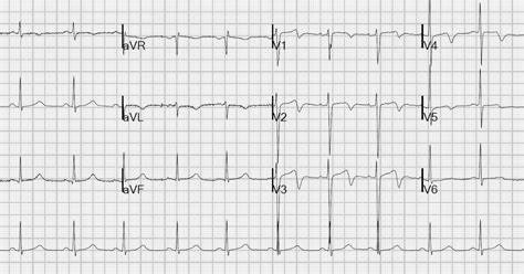
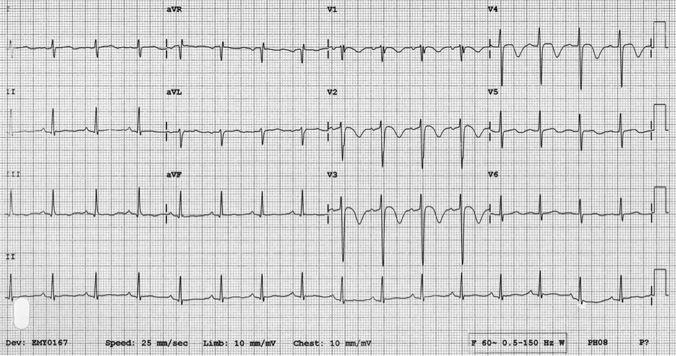
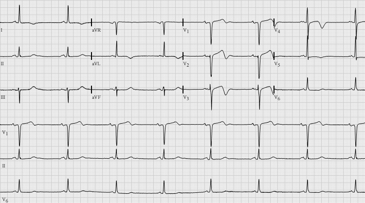
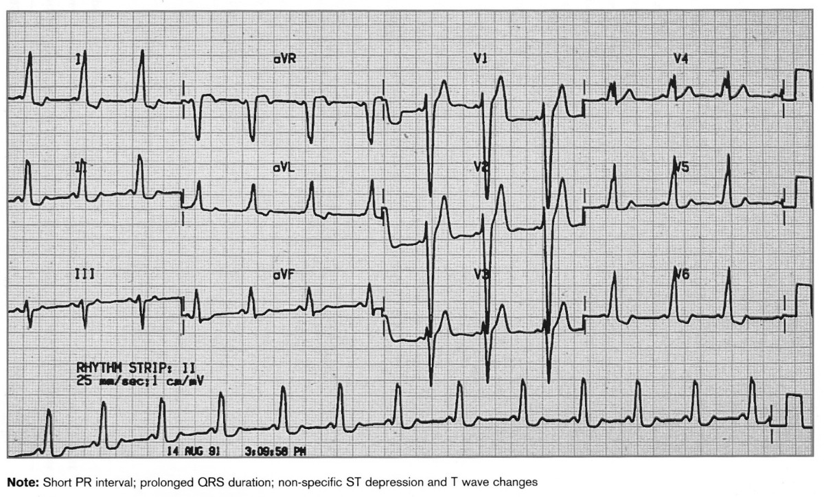
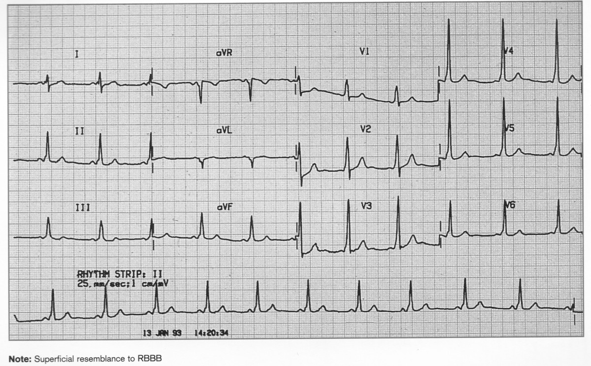
WPW
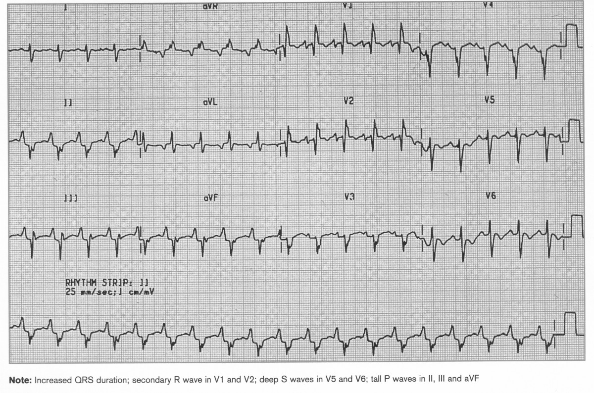
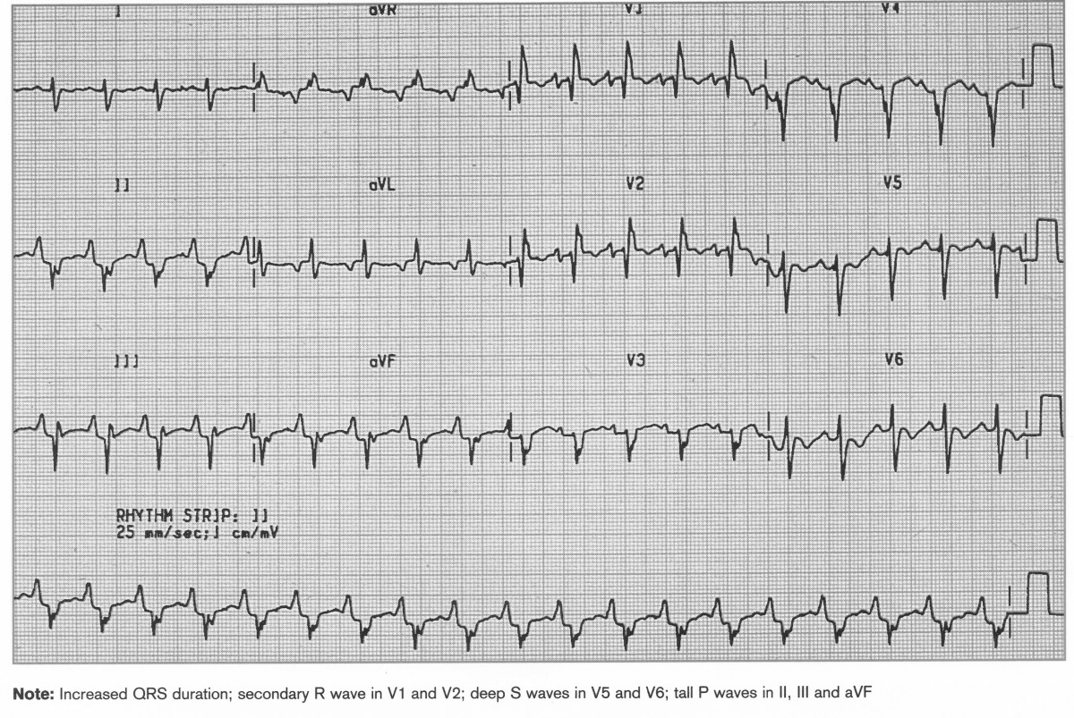
RVH
Increased QRS duration, secondary R wave in V1 and V2, deep S V5V6 tall P waves in II,III,aVF
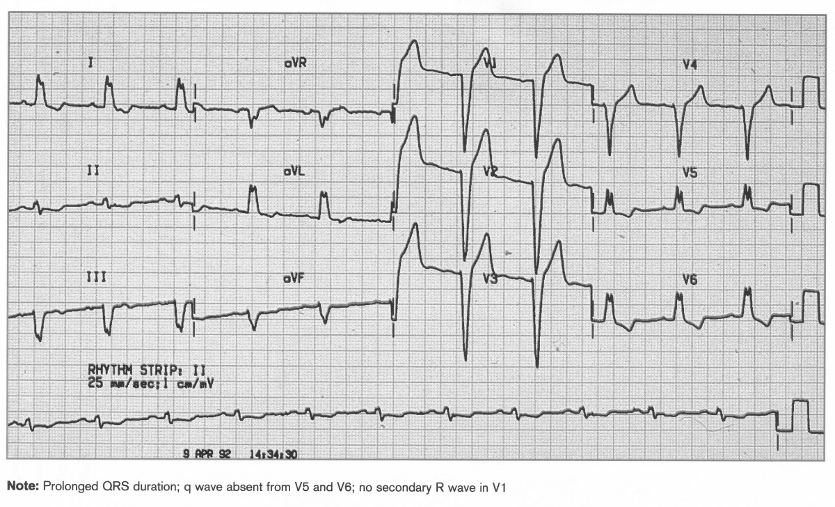
Left Bundle Branch Block
Prolonged QRS duration, q wave absent V5 and V6, no secondary R wave in V1
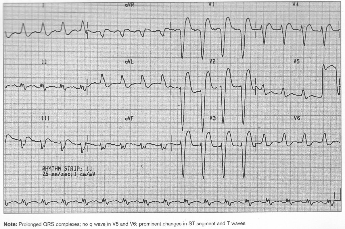
Prolonged QRS, no q wave in V5 and V6, prominent changes in ST segment and T wave
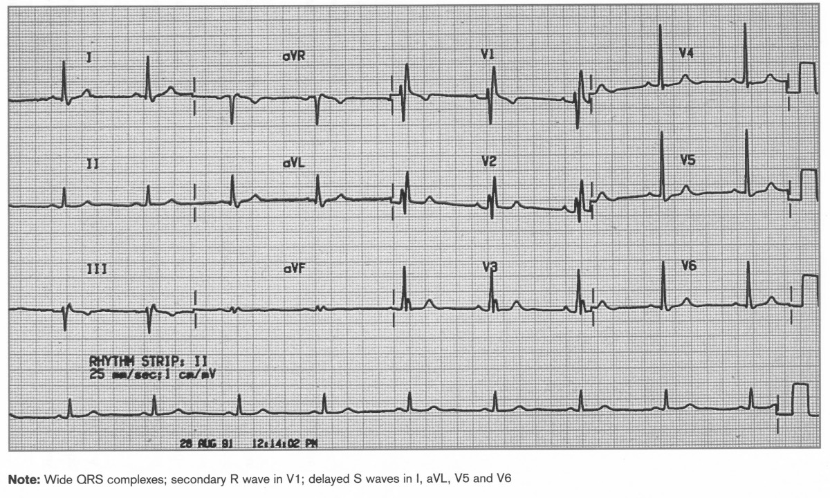
Right Bundle Branch Block
Wide QRS, secondary R wave in V1 and delayed S waves in I, aVL, V5 and V6
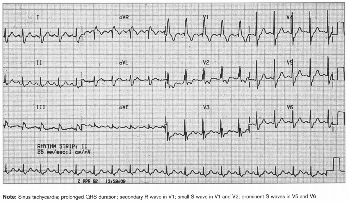
Sinus tachycardia, prolonged QRS duration, secondary R wave in V1 and V2, prominent S waves in V5 and V6>
IHD
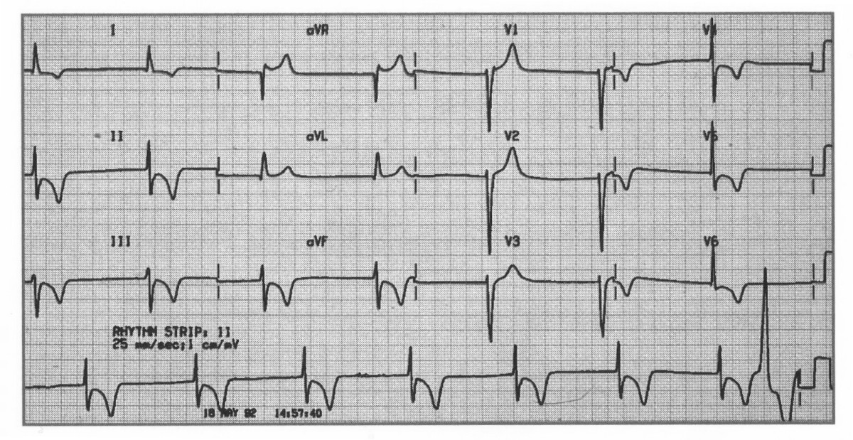
Recent Anterolateral MI
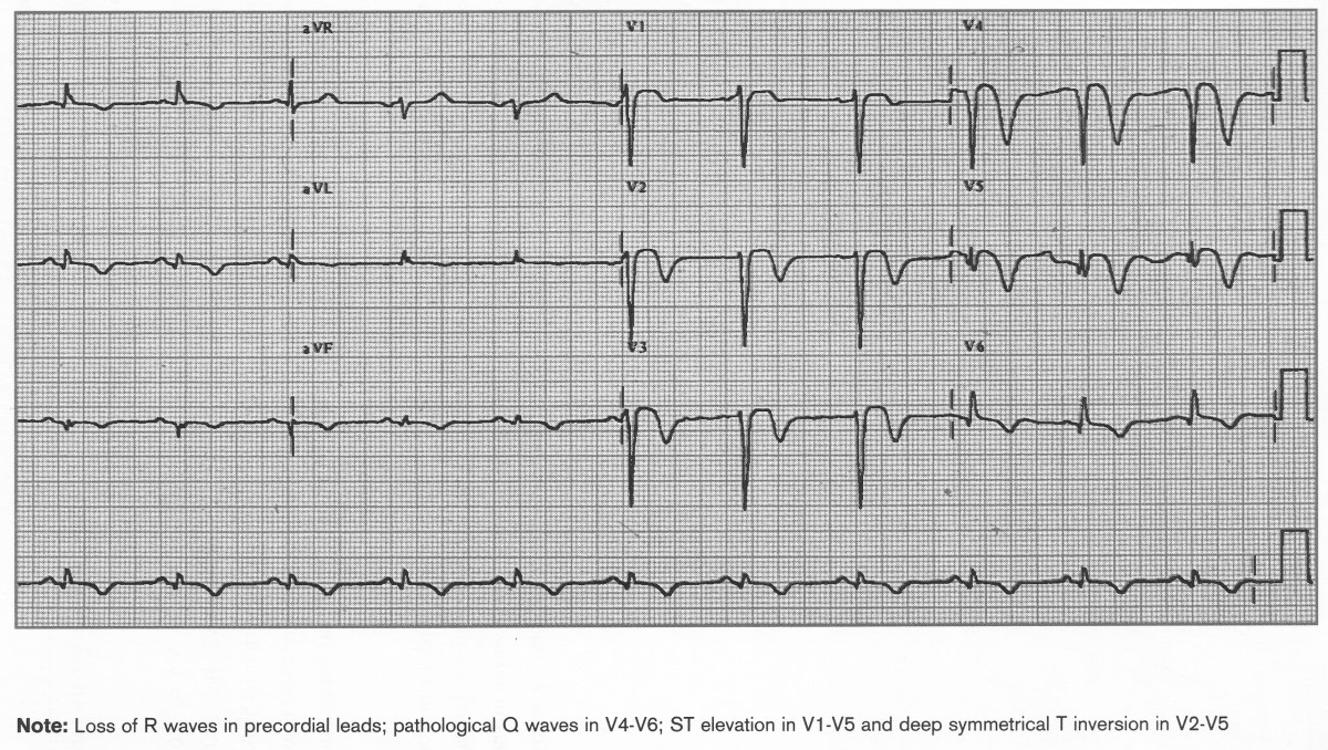
Acute Inferior MI, Old Anteroseptal MI
ST elevation II,III,aVF, ST depression and T wave inversion I,aVL, V2-V5. Poor R wave progression
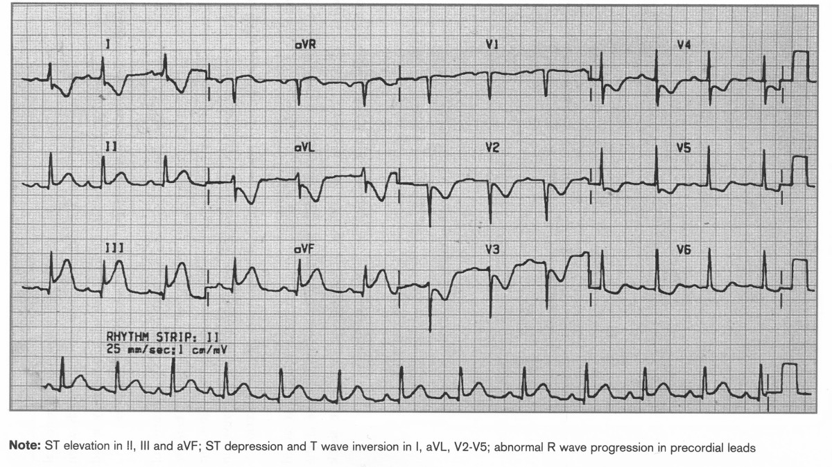
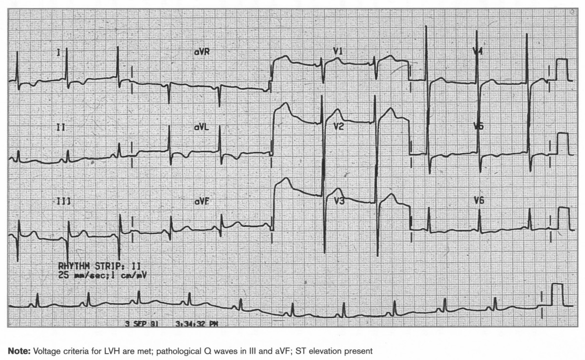
Acute Anterior STEMI, old inferior MI
ST elevation, no R waves V1-V6, Pathological Q waves in III and aVF
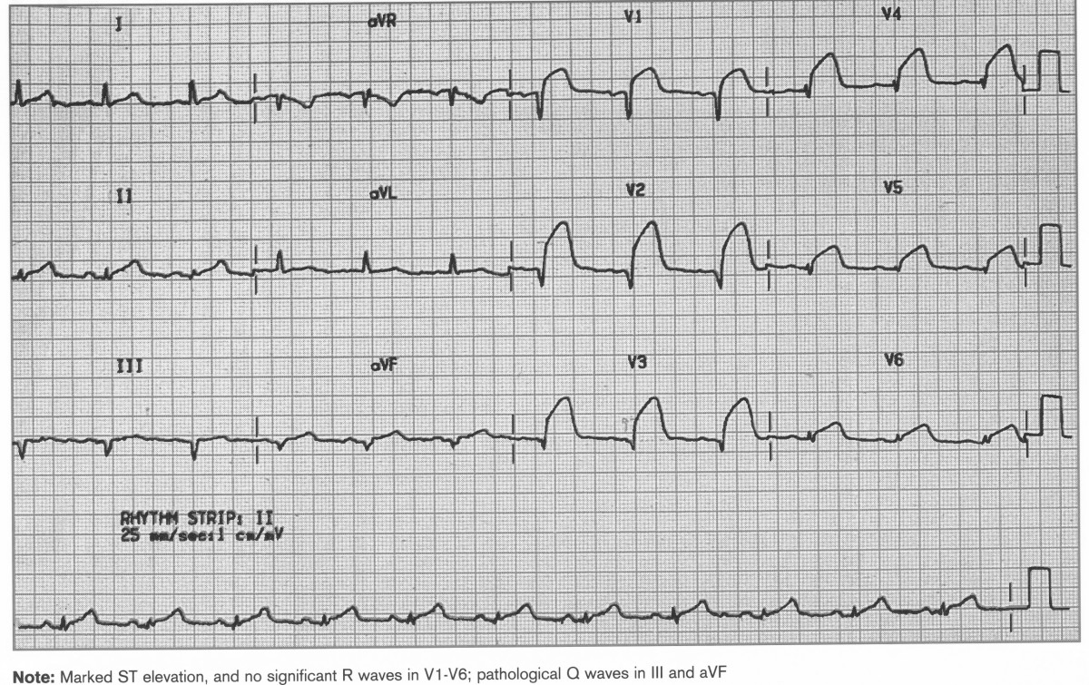
???Q waves V1-V5, lack of R wave in precordial leads, ST elevation V2-V5, T waves inverted I, aVL, V2-V5
Recent Anterior-lateral STEMI
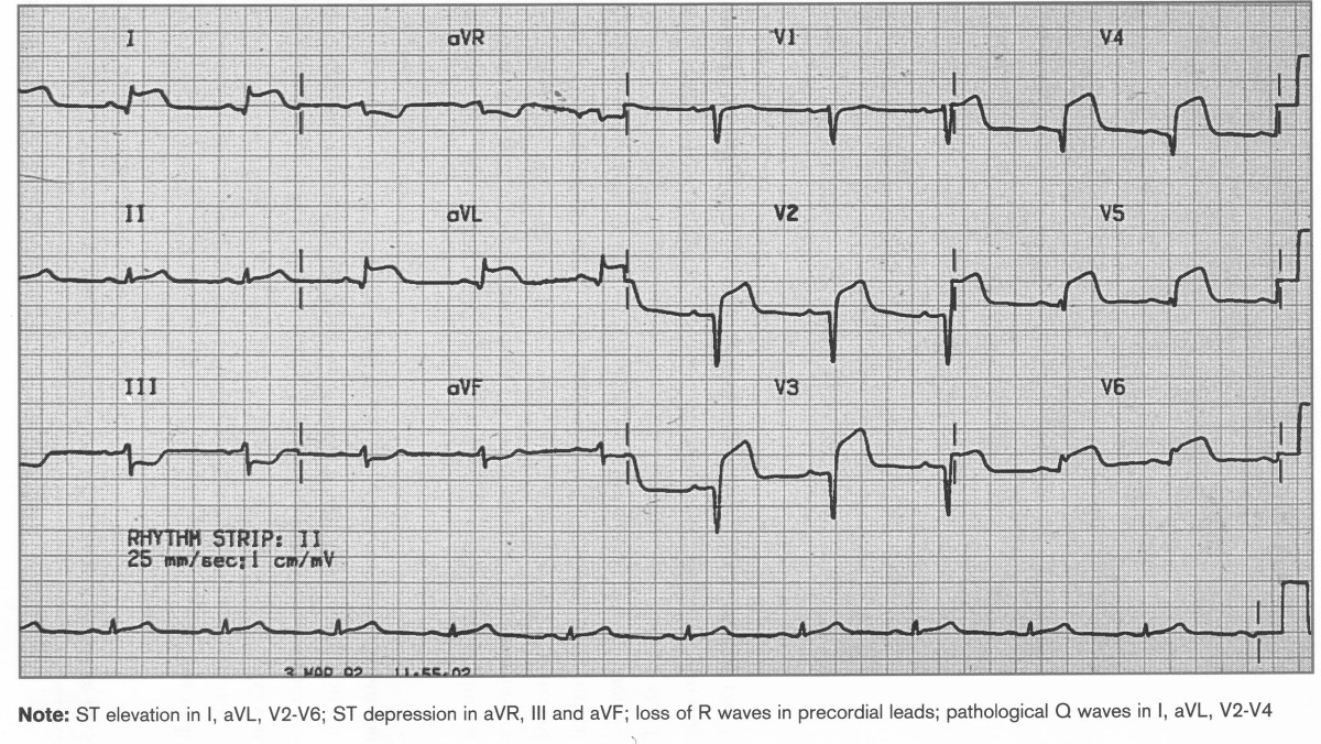
ST elevation I, aVL, V2-V6, ST depression a VR, III, aVF. Loss of R waves, path Q waves I,aVL, V2-V4
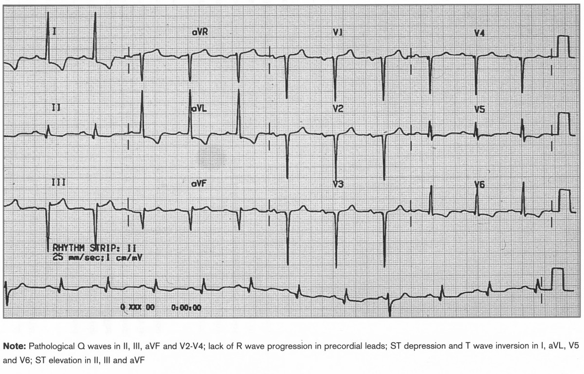
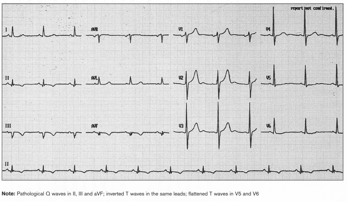
Old Inferior STEMIM
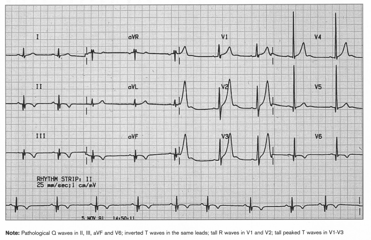
Pathological Q waves in II, III, aVF with inverted T waves. Flattened T waves in V5=V6
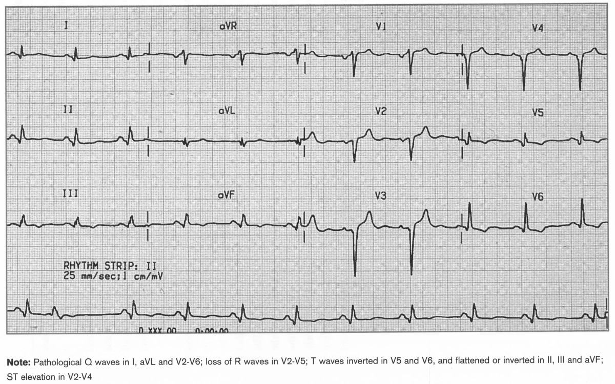
Old Anterolateral MI
Pathological Q waves in I,aVl, V2-6, Loss of R waves V2-5, T waves inverted V5-6 and flattened or inverted in II,III, avF, ST elevation V2-4
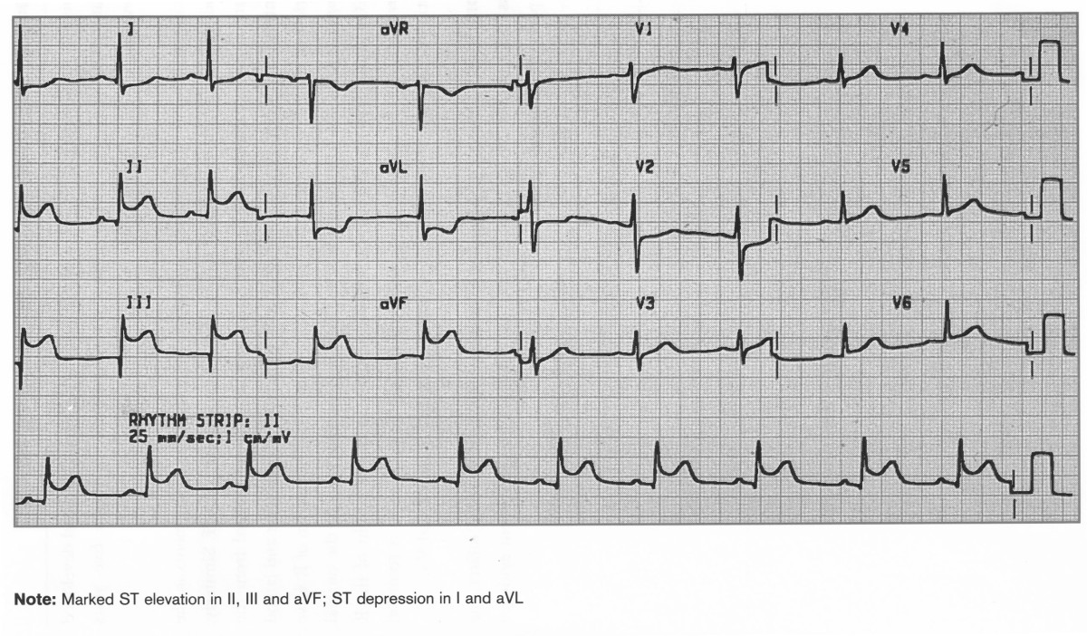
Acute Inferior STEMI
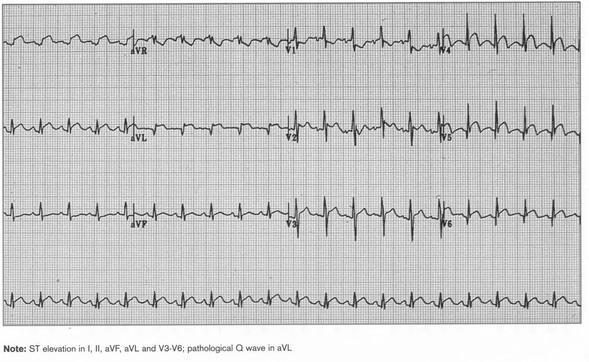
Acute Inferolateral MI
ST elevation I, II, aVF, aVL, V3-V6, Pathological Q wave in aVL

Acute Inf MI and True Posterior
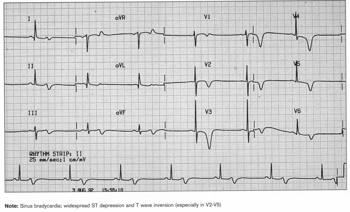
NSTEMI
Sinus bradycardia, ST depression widespread, T wave inversion V2-5
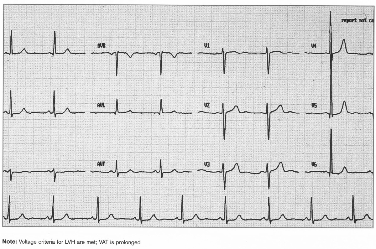
Hypertension
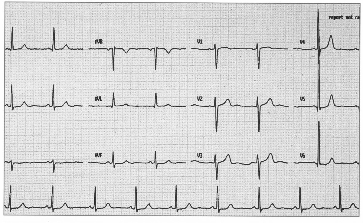
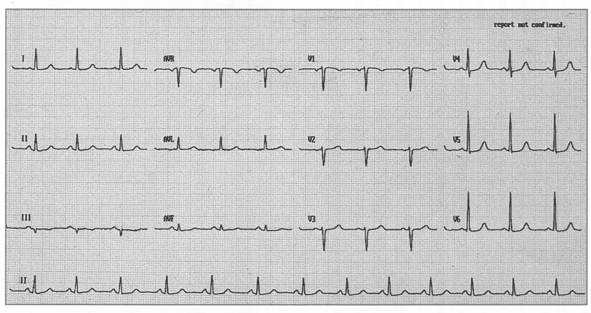
Voltage criteria for LVH are met, Ventricular activation time is prolonged
Voltage criteria for LVH are met, pathological Q waves in III and aVf and ST elevation suggest LVH and recent Inferior MI. Ventricular activation time is prolonged
