Mycosis Fungoides (Sezary Syndrome)
Related Subjects:
|Nikolsky's sign
|Koebner phenomenon
|Erythema Multiforme
|Pyoderma gangrenosum
|Erythema Nodosum
|Dermatitis Herpetiformis
|Lichen Planus
|Acanthosis Nigricans
|Acne Rosacea
|Acne Vulgaris
|Alopecia
|Vitiligo
|Urticaria
|Basal Cell Carcinoma
|Malignant Melanoma
|Squamous Cell Carcinoma
|Mycosis Fungoides (Sezary Syndrome)
|Xeroderma pigmentosum
|Bullous Pemphigoid
|Pemphigus Vulgaris
|Seborrheic Dermatitis
|Pityriasis/Tinea versicolor infections
|Pityriasis rosea
|Scabies
|Dermatomyositis
|Toxic Epidermal Necrolysis
|Stevens-Johnson Syndrome
|Atopic Eczema/Atopic Dermatitis
|Psoriasis
📖 About
- Mycosis fungoides is a rare cutaneous T-cell lymphoma (CTCL), and the most common subtype.
- It manifests primarily on the skin with multiple erythematous, itchy lesions, often mistaken for eczema or psoriasis → delayed diagnosis.
- Usually progresses slowly 🐢 and may not shorten life expectancy in early/limited disease.
- When malignant T-cells circulate in blood with systemic involvement → classified as Sézary Syndrome (aggressive form 🔴).
🧬 Aetiology
- A neoplastic proliferation of CD4+ T-helper lymphocytes with skin-homing properties.
- Typically affects adults aged 40–60 years, with a slight male predominance.
- Exact cause unknown, but chronic antigenic stimulation, genetic factors, and immune dysregulation are implicated.
👀 Clinical Features
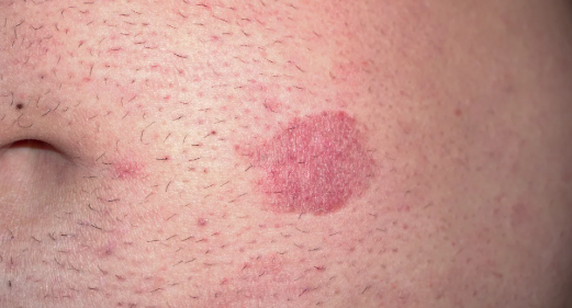
- Chronic, itchy, erythematous patches → slowly progress over years.
- Early lesions: scaly, irregular patches on sun-protected areas (buttocks, thighs, trunk).
- Can mimic psoriasis/eczema, delaying diagnosis ⏳.
- Later: plaques, nodules, ulcerated tumours (may discharge).
- Diffuse redness of skin → erythroderma.
- Advanced: exfoliative dermatitis, generalised lymphadenopathy, and ~10% risk of visceral organ involvement (liver, spleen, lung).
- Sézary syndrome: erythroderma + lymphadenopathy + malignant T-cells in blood.
🔬 Investigations
- Skin biopsy (diagnostic): infiltrate of atypical CD4+ T-cells with cerebriform (Sézary-Lutzner) nuclei; epidermotropism with Pautrier’s microabscesses.
- Blood tests: usually normal in early disease; check for systemic spread.
- Flow cytometry if Sézary syndrome suspected (circulating malignant T-cells).
- Staging workup: CT/PET to assess nodal/visceral involvement.
📊 Staging (simplified)
- Patch stage – erythematous scaly patches (years to decades).
- Plaque stage – thickened, raised lesions.
- Tumour stage – nodules, ulcerating masses.
- Systemic stage – blood, lymph node, organ involvement (Sézary syndrome).
💊 Management
- No absolute cure, but remission and long-term control possible.
- Early disease often treated with skin-directed therapies:
- Regular emollients + topical corticosteroids for itch/inflammation.
- Phototherapy (NB-UVB or PUVA) for widespread skin involvement.
- Local radiotherapy (electron beam, low-voltage X-rays) for thick plaques/tumours.
- For refractory or advanced disease → systemic therapies:
- Methotrexate – immunosuppressive, first-line systemic agent.
- Oral retinoids – normalise keratinocyte growth.
- Interferon-alpha – immune modulation.
- Extracorporeal photopheresis – esp. in Sézary syndrome.
- Chemotherapy – reserved for aggressive/refractory disease (e.g., CHOP regimen).
- Care is MDT-led 👥 → dermatology, haematology, oncology input.
✅ Key Exam Pearls
- Most common cutaneous T-cell lymphoma.
- Biopsy shows cerebriform CD4+ cells + epidermotropism.
- Sézary syndrome = triad (erythroderma + lymphadenopathy + circulating malignant cells).
- Treatment is stage-based → skin therapies early, systemic if advanced.
