
| Download the amazing global Makindo app: ✅ Means NICE/National Guidelines 2026 compliant Android | Apple | |
|---|---|
| MEDICAL DISCLAIMER: Educational use only. Not for diagnosis or management. See below for full disclaimer. |
Triangles of the neck
Related Subjects: |Neck Swellings by Triangle |Thyroglossal cyst |Branchial cleft cyst |Head and Neck Cancers |Triangles of the neck
📖 About
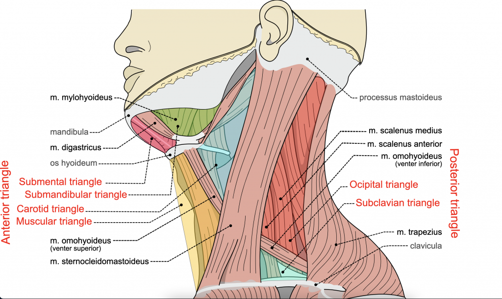
- The triangles of the neck are surface anatomical regions formed by the sternocleidomastoid (SCM) and other boundaries, divided into an anterior triangle and a posterior triangle.
- These triangles contain vital neurovascular and visceral structures, making them clinically important in surgery, trauma, and examination.
🔺 Anterior Triangle Borders
- Superior: Inferior border of the mandible 🦷
- Medial: Midline of the neck
- Lateral: Anterior border of the SCM
📌 Subdivisions of the Anterior Triangle
- Muscular (Omotracheal) Triangle: Contains infrahyoid muscles, thyroid & parathyroids 🦋
- Carotid Triangle: Carotid artery, IJV, vagus nerve – 💉 common for carotid endarterectomy
- Submandibular Triangle: Submandibular gland, facial artery/vein, CN XII
- Submental Triangle: Submental lymph nodes – 🔎 first site for oral cancer spread
🔹 Posterior Triangle Borders
- Anterior: Posterior margin of SCM
- Posterior: Anterior border of trapezius
- Inferior: Middle one-third of clavicle
📌 Subdivisions of the Posterior Triangle
- Occipital Triangle: Contains accessory nerve (CN XI) – ⚠️ vulnerable in lymph node biopsy
- Supraclavicular (Omoclavicular) Triangle: Subclavian artery/vein, brachial plexus roots – 🚑 important in central line insertion and trauma
💡 Clinical Relevance
- Swelling in the anterior triangle often suggests thyroid or lymph node pathology.
- Posterior triangle lumps are more likely lymphadenopathy (e.g., lymphoma, metastasis).
- Knowledge of these landmarks is crucial for surgical approaches (e.g., carotid surgery, neck dissections).
📚 References
| The content on this website is provided for educational and informational purposes only to support exam preparation (e.g., MLA, MRCP, USMLE) and learning. This is NOT medical advice, diagnosis, treatment, or professional guidance. It does not replace consultation with a qualified healthcare professional, official guidelines (e.g., NICE, GMC, BNF), or supervised clinical practice. Always verify information with current, authoritative sources. Makindo and its contributors accept no liability for any reliance on this content, including errors, omissions, or any resulting harm, loss, or consequences. By using this site, you agree to these terms. |
|
|
Categories
- About
- Acute Medicine
- Anaesthetics and Critical Care
- Anatomy
- Anatomy and Physiology
- Biochemistry
- Book
- Cardiology
- Collections
- CompSci
- Crib Sheets
- Critical care
- Dental
- Dermatology
- Differentials
- Drugs
- ENT
- Electrocardiogram
- Embryology
- Emergency Medicine
- Endocrinology
- Ethics
- Foundation Doctors
- GCSE
- Gastroenterology
- General Practice
- Genetics
- Geriatric Medicine
- Geriatrics
- Guidelines
- Haematology
- Hepatology
- Immunology
- Infectious Diseases
- Infographic
- Investigations
- Lists
- MRCP
- Mandatory Training
- Medical Students
- Microbiology
- Nephrology
- Neurology
- Neurosurgery
- Nutrition
- OSCE
- Obstetrics Gynaecology
- Oncology
- Ophthalmology
- Oral Medicine and Dentistry
- Orthopaedics
- Paediatrics
- Palliative
- Palliative Care
- Pathology
- Pharmacology
- Physiology
- Procedures
- Psychiatry
- Public Health
- Radiology
- Respiratory
- Resuscitation
- Revision Topics
- Rheumatology
- Statistics and Research
- Stroke
- Surgery
- Toxicology
- Trauma and Orthopaedics
- USMLE
- Urology
- Vascular Surgery