Makindo Medical Notes"One small step for man, one large step for Makindo" |
|
|---|---|
| Download all this content in the Apps now Android App and Apple iPhone/Pad App | |
| MEDICAL DISCLAIMER: The contents are under continuing development and improvements and despite all efforts may contain errors of omission or fact. This is not to be used for the assessment, diagnosis, or management of patients. It should not be regarded as medical advice by healthcare workers or laypeople. It is for educational purposes only. Please adhere to your local protocols. Use the BNF for drug information. If you are unwell please seek urgent healthcare advice. If you do not accept this then please do not use the website. Makindo Ltd. |
Basic Neuroscience
-
| About | Anaesthetics and Critical Care | Anatomy | Biochemistry | Cardiology | Clinical Cases | CompSci | Crib | Dermatology | Differentials | Drugs | ENT | Electrocardiogram | Embryology | Emergency Medicine | Endocrinology | Ethics | Foundation Doctors | Gastroenterology | General Information | General Practice | Genetics | Geriatric Medicine | Guidelines | Haematology | Hepatology | Immunology | Infectious Diseases | Infographic | Investigations | Lists | Microbiology | Miscellaneous | Nephrology | Neuroanatomy | Neurology | Nutrition | OSCE | Obstetrics Gynaecology | Oncology | Ophthalmology | Oral Medicine and Dentistry | Paediatrics | Palliative | Pathology | Pharmacology | Physiology | Procedures | Psychiatry | Radiology | Respiratory | Resuscitation | Rheumatology | Statistics and Research | Stroke | Surgery | Toxicology | Trauma and Orthopaedics | Twitter | Urology
Related Subjects: |Brain Herniation syndromes |Haemorrhagic stroke |Traumatic Head/Brain Injury |Acute Hydrocephalus |Epidural Haematoma |Subdural haematoma |Basic Neuroscience |Medulla Oblongata |Midbrain |Pons |Caudate Nucleus |Putamen and Globus Pallidus |Cerebral Cortex |Internal Capsule |Cavernous sinus |Basal Ganglia
🧠 Cells in the CNS
The brain contains ~1011 neurons and even more glial cells (outnumbering neurons 10–50×). 🧬 About 40% of the human genome contributes to brain development & function. ⚡ Neurons rely on continuous ATP from oxidative phosphorylation → brain is highly metabolic.
CNS = neurons + glial cells. Both form the neurovascular unit, essential in stroke pathophysiology.
🧩 Neuronal Cells
Neurons = “information processors” → receive input, integrate, and send output signals. Grey matter = soma clusters (cortex, nuclei, brainstem, cerebellum).
Key parts:
- 🟢 Soma: nucleus & organelles.
- 🌳 Dendrites: receive input.
- ⚡ Axon hillock: AP initiation.
- ➡️ Axon: conducts impulse.
- 📤 Terminals: release neurotransmitters at synapses.
🔄 Transport: - Anterograde → soma → terminal. - Retrograde → terminal → soma (used by rabies, tetanus).
 🧪 Non-Neuronal Cells (Glia)
Glia = “glue” but vital support + barrier + repair functions:
- 🌟 Astrocytes: scaffold, ion buffering, neurotransmitter recycling, maintain BBB.
- 🛡️ Microglia: immune cells, phagocytose debris & pathogens, regulate inflammation.
- 💧 Ependymal cells: line ventricles, circulate CSF, tumour origin (ependymoma).
- 🧵 Oligodendrocytes: myelinate CNS axons → saltatory conduction. One cell → many axons.
⚡ Cell Electrophysiology
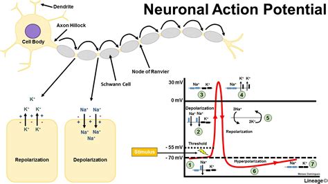
- Resting potential: ~ –70 mV (Na⁺/K⁺ pump + selective permeability).
- AP: Na⁺ influx → depolarisation; K⁺ efflux → repolarisation.
- Myelination → saltatory conduction at nodes of Ranvier. ❌ Lost in MS → conduction slowed.
 🔄 Synapses, Neurotransmitters & Receptors
- 🔥 Glutamate: excitatory (NMDA, AMPA).
- 🛑 GABA: inhibitory (GABAA, GABAB).
- ⚡ Acetylcholine: nicotinic (ionotropic), muscarinic (metabotropic).
- 🎭 Monoamines: dopamine, serotonin, norepinephrine → mood, cognition, arousal.
🧠 Synaptic plasticity (LTP/LTD) = basis of learning & memory.
💓 Brain Physiology & Cerebral Perfusion
- Brain = 1–2% body weight but needs 15% cardiac output + 20% O₂.
- Metabolism: glucose oxidation, minimal reserves.
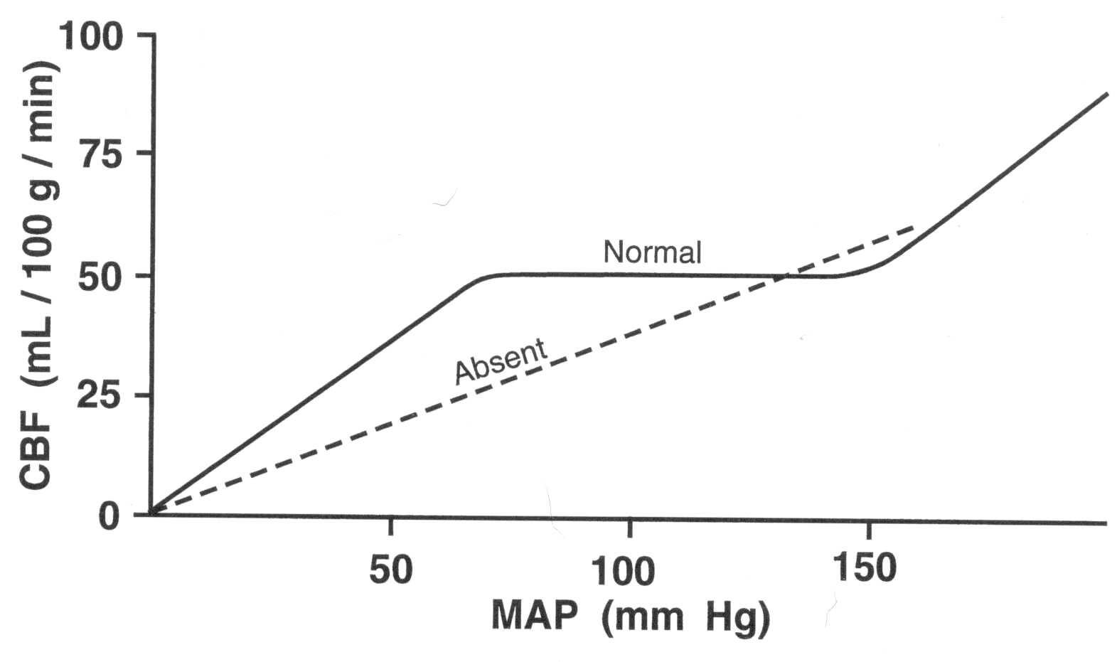
- Autoregulation adjusts flow to BP, CO₂, pH → fails in stroke.
 🩸 Stroke Pathophysiology
Sudden interruption of cerebral blood flow → neuronal death.
| 🧠 Three Zones Post-Stroke | |
|---|---|
| ☠️ Irreversible core | Necrosis from Ca²⁺ influx, osmotic failure. |
| 🟡 Penumbra | Salvageable if reperfused quickly. |
| 🟢 Mild hypoperfusion | Viable but impaired; oxidative stress risk. |
⏱️ Time is brain – reperfusion (thrombolysis/thrombectomy) saves the penumbra. MRI-DWI distinguishes core vs at-risk tissue.
📊 Factors Influencing Stroke Outcome
- ⏳ Duration/speed of ischaemia (slower = more adaptation).
- 🌐 Collateral circulation.
- 🩺 Systemic BP and perfusion pressure.
- 🩸 Clotting tendency & fibrinolysis.
- 🌡️ Hyperthermia worsens damage.
- 🍬 Hyperglycaemia = larger infarcts, worse prognosis.