
| Download the amazing global Makindo app: ✅ Means NICE/National Guidelines 2026 compliant Android | Apple | |
|---|---|
| MEDICAL DISCLAIMER: Educational use only. Not for diagnosis or management. See below for full disclaimer. |
Ehlers-Danlos syndromes
Related Subjects: |Autosomal Dominant |Autosomal Recessive |X Linked Recessive
🌟 Ehlers-Danlos Syndromes (EDS) are a group of inherited connective tissue disorders caused by defects in collagen synthesis and structure. Collagen is the scaffolding protein of skin, joints, vessels, and organs-so when it is faulty, the entire support system of the body is weakened. Common hallmarks include 🤸 joint hypermobility, 🧵 skin hyperextensibility, and 💥 tissue fragility, but the specific presentation varies across subtypes.
📖 About
- A heterogeneous collection of conditions, historically classified as EDS I–X, now recognized as 13 subtypes.
- Overall prevalence ~1 in 5,000 births 👶.
- Most forms are autosomal dominant, though some are autosomal recessive.
- EDS represents a spectrum: some mild with joint laxity, others life-threatening with vascular rupture.
🧬 Aetiology
- Mutations in genes encoding collagen or enzymes that process collagen (e.g., COL5A1, COL3A1, PLOD1).
- Defective collagen biosynthesis → abnormal fibrils → weak connective tissue.
- Faults in post-translational modification (e.g., hydroxylation, cross-linking) impair tensile strength.
- Take-home: study collagen pathways-EDS is essentially “molecular scaffolding gone wrong.”
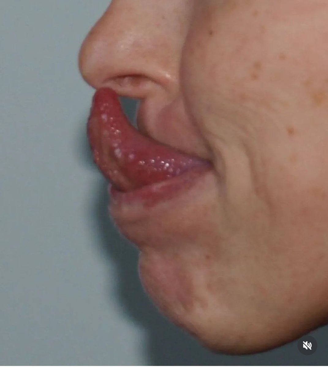
Gorlin’s sign 👅
🔎 Types of Ehlers-Danlos Syndromes
There are 13 recognized subtypes; the most important exam-relevant ones include:
- Hypermobile EDS (hEDS) 🤸: Most common; marked joint hypermobility, chronic pain, soft/velvety skin. Genetics still not fully defined.
- Classical EDS (cEDS) 🧵: Soft, hyperelastic skin, widened atrophic scars, frequent bruising, joint laxity. Mutations in COL5A1/2.
- Vascular EDS (vEDS) 💔: Most dangerous; fragile arteries, risk of rupture, bowel and uterine rupture. Associated with COL3A1 mutation.
- Kyphoscoliotic EDS (kEDS) 🌀: Severe congenital scoliosis, hypotonia, ocular fragility. Caused by PLOD1 mutations (lysyl hydroxylase deficiency).
- Arthrochalasia EDS (aEDS) 🦴: Severe congenital hip dislocations, extreme joint hypermobility, fragile skin.
- Dermatosparaxis EDS (dEDS) 🩹: Extremely fragile, sagging skin; hernias. Caused by ADAMTS2 mutation.
🩺 Clinical Features
- 🤸 Joint hypermobility: “party trick” joints but prone to sprains/dislocations.
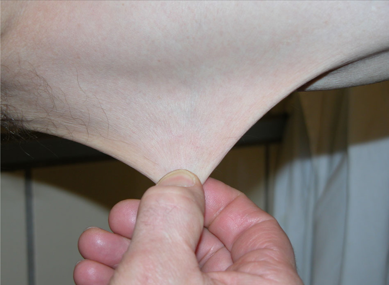
- 🧵 Skin hyperextensibility: Soft, stretchy skin, sometimes with cigarette-paper scars.
- 💥 Tissue fragility: Easy bruising, poor wound healing, atrophic scars.
- 🩹 Musculoskeletal: Chronic pain, fatigue, frequent dislocations, scoliosis.
- 👁️ Ocular: Fragile sclerae → risk of globe rupture (esp. in kEDS).
- ❤️ Cardiovascular: Aortic dilatation, mitral valve prolapse, vascular rupture in vEDS.
- ⚠️ GI: Bowel rupture, hernias.
🧾 Differentials
- Familial Joint Hypermobility (benign, no tissue fragility)
- Marfan’s Syndrome (tall habitus, lens dislocation, fibrillin mutation)
- Larsen’s Syndrome (joint dislocations, craniofacial features)
🧪 Investigations
- Clinical criteria: Beighton score for hypermobility, skin/vascular signs.
- Family history: Key to inheritance pattern.
- Genetic testing: Confirms subtype (esp. vEDS via COL3A1).
- Imaging: Echocardiography for aortic root; MRI for scoliosis/joint pathology.
- Skin biopsy: Can show abnormal collagen fibrils (research/rarely clinical).
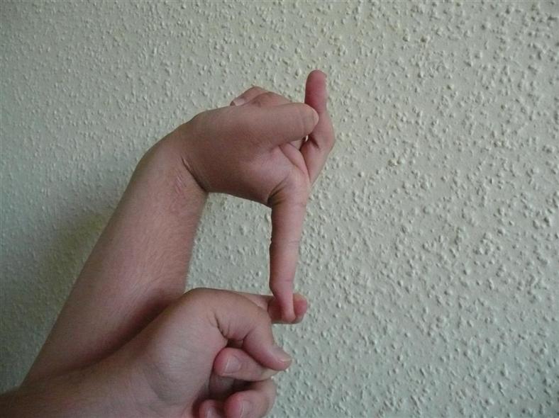
Beighton score (0–9) 🦴
The Beighton score is a quick bedside screen for generalised joint hypermobility. It scores 5 manoeuvres (4 are bilateral) to give a total out of 9 - it supports (but does not diagnose) hypermobility spectrum disorders / hEDS. Hypermobility reflects increased connective-tissue laxity (collagen/ECM mechanics), so pair the score with symptoms (pain, sprains, dislocations, dysautonomia features) and skin/family history.
- 1 point each side: Passive dorsiflexion of the 5th finger > 90° (R / L)
- 1 point each side: Thumb touches the forearm (R / L)
- 1 point each side: Elbow hyperextension > 10° (R / L)
- 1 point each side: Knee hyperextension > 10° (R / L)
- 1 point: Palms flat on the floor with knees straight (forward flexion)
- Common cut-offs: ≥ 5/9 adults, ≥ 6/9 children/adolescents, ≥ 4/9 if >50 years (thresholds vary by guideline/context).
- Practical tip: score what you see today (don’t “award for history”), but note pain, prior surgery, and guarding can reduce apparent hypermobility.
💊 Management
- Physiotherapy 🤸: Strengthen periarticular muscles; prevent dislocations.
- Pain management 💊: Paracetamol, NSAIDs, avoid long-term opioids; sometimes neuropathic pain agents.
- Protective strategies 🦾: Braces, taping, mobility aids, avoiding high-impact sports.
- Cardiac surveillance ❤️: Especially in vEDS (yearly echo, vascular imaging).
- Surgery 🩺: High risk due to fragile tissues-reserved for emergencies or severe complications.
- Genetic counseling 🧬: For patients and families.
📌 Clinical Pearls
- Not every hypermobile patient has EDS-distinguish from benign hypermobility.
- vEDS = ⚠️ think “vascular catastrophe” → young person with spontaneous arterial rupture, uterine rupture, or bowel perforation.
- EDS scars often look like “cigarette paper” 🚬-thin, papery, and stretched.
- Always screen for psychological burden-chronic pain & fatigue are disabling.
| The content on this website is provided for educational and informational purposes only to support exam preparation (e.g., MLA, MRCP, USMLE) and learning. This is NOT medical advice, diagnosis, treatment, or professional guidance. It does not replace consultation with a qualified healthcare professional, official guidelines (e.g., NICE, GMC, BNF), or supervised clinical practice. Always verify information with current, authoritative sources. Makindo and its contributors accept no liability for any reliance on this content, including errors, omissions, or any resulting harm, loss, or consequences. By using this site, you agree to these terms. |
|
|
Categories
- About
- Acute Medicine
- Anaesthetics and Critical Care
- Anatomy
- Anatomy and Physiology
- Biochemistry
- Book
- Cardiology
- Collections
- CompSci
- Crib Sheets
- Critical care
- Dental
- Dermatology
- Differentials
- Drugs
- ENT
- Electrocardiogram
- Embryology
- Emergency Medicine
- Endocrinology
- Ethics
- Foundation Doctors
- GCSE
- Gastroenterology
- General Practice
- Genetics
- Geriatric Medicine
- Geriatrics
- Guidelines
- Haematology
- Hepatology
- Immunology
- Infectious Diseases
- Infographic
- Investigations
- Lists
- MRCP
- Mandatory Training
- Medical Students
- Microbiology
- Nephrology
- Neurology
- Neurosurgery
- Nutrition
- OSCE
- Obstetrics Gynaecology
- Oncology
- Ophthalmology
- Oral Medicine and Dentistry
- Orthopaedics
- Paediatrics
- Palliative
- Palliative Care
- Pathology
- Pharmacology
- Physiology
- Procedures
- Psychiatry
- Public Health
- Radiology
- Respiratory
- Resuscitation
- Revision Topics
- Rheumatology
- Statistics and Research
- Stroke
- Surgery
- Toxicology
- Trauma and Orthopaedics
- USMLE
- Urology
- Vascular Surgery