Related Subjects:
|Causes of abnormal Vaginal bleeding
|Vaginal Carcinoma
|Cervical cancer
|Cervical cancer screening
|Antepartum haemorrhage
|Trauma: Traumatic Brain Head Injury (TBI)
|Post Menopausal Bleeding
|Postpartum haemorrhage
🧪 Cervical Cancer Screening helps detect precancerous changes early, allowing treatment before progression to invasive cancer.
It involves sampling cells from the cervix and analysing them for abnormalities.
Early detection → better outcomes ✅
📌 About
- Cells collected from the ectocervix using a spatula or brush.
- Sample placed on a glass slide or in liquid-based cytology medium → stained & examined in the lab.
- Goal: Detect precancerous lesions early → prevent progression to cervical cancer.
📅 Screening Guidelines (UK)
- 👩 Women aged 25–49 years: Invited every 3 years.
- 👩🦳 Women aged 50–64 years: Invited every 5 years.
- Women >65: Only screened if not tested since 50 or if recent abnormal results.
- Screening may be modified if high risk (e.g., HIV, immunosuppression).
🧾 Pap Smear Results
- Mild Dyskaryosis: Often due to transient HPV infection; usually low risk.
- Moderate Dyskaryosis: More significant abnormalities; likely CIN 2/3.
- Severe Dyskaryosis: High-grade abnormality → high risk of progression to cancer.
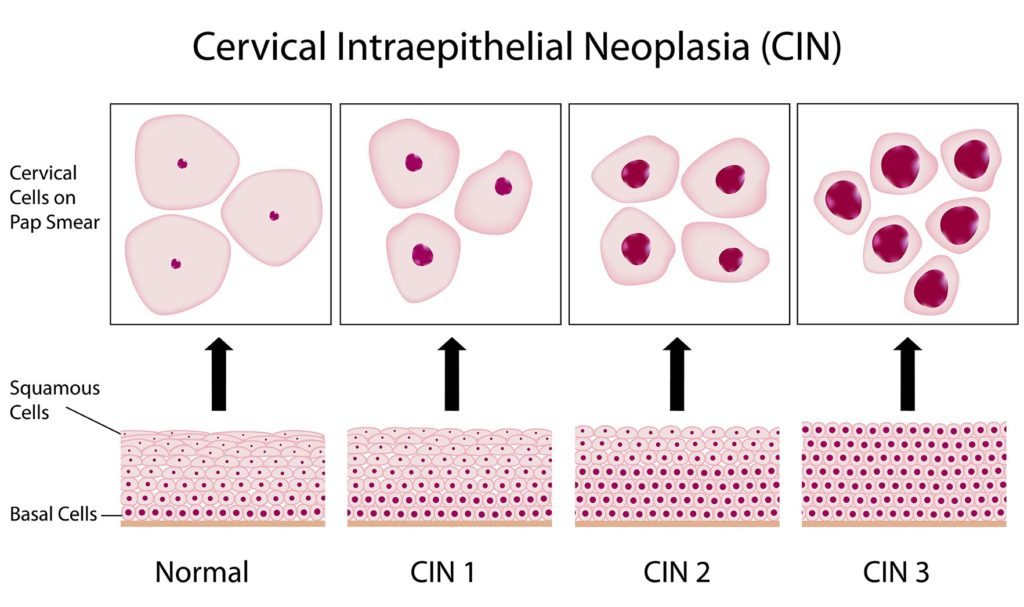
🔬 Cervical Intraepithelial Neoplasia (CIN)
- CIN 1: Abnormal cells in lower third of epithelium → low-grade; many regress spontaneously.
- CIN 2: Abnormal cells in two-thirds of epithelium → high-grade; usually needs treatment.
- CIN 3: Abnormal cells throughout full epithelial thickness → high risk; requires treatment.
⚡ Actions Based on Results
- Mild dyskaryosis / CIN 1: Repeat smear in 6–12 months or colposcopy referral.
- Moderate dyskaryosis / CIN 2: Refer for colposcopy + likely treatment.
- Severe dyskaryosis / CIN 3: Urgent colposcopy + excisional treatment (e.g. LLETZ).
🩺 Management of Precancerous Changes
- Colposcopy: Magnified cervix examination ± biopsy; treatment includes laser ablation, cold coagulation, or cryotherapy.
- LLETZ / Cone biopsy: Removes transformation zone containing abnormal cells.
🔑 Cone biopsy reserved for glandular disease or unclear margins.
- Follow-up: Repeat smear/HPV test at 6 months → ensure clearance.
ℹ️ General Management Notes
- Benign findings (e.g., metaplasia, atrophic smear) → no action.
- Inflammatory changes → test for infections (e.g., chlamydia, candida, bacterial vaginosis).
- Actinomyces on smear with IUCD → consider IUCD removal + gynae review.
- Glandular neoplasia: Always urgent referral (risk of adenocarcinoma).
📚 References
