HIV patients are at increased risk of infections like Strep pneumoniae, Haemophilus influenzae, and Moraxella catarrhalis.
About
- Pneumocystis carinii was renamed Pneumocystis jirovecii, named after the physician who first described the organism as a cause of pneumonia in neonates. Despite the name change, the term PCP (Pneumocystis pneumonia) remains commonly used.
- Pneumocystis jirovecii is a fungal organism lacking ergosterol, making it resistant to conventional antifungal drugs.
Aetiology
- Occurs in immunocompromised patients, especially those with HIV/AIDS and a CD4 count < 200 cells/mm3.
- May be seen at initial presentation of HIV or in those who have stopped antiretroviral therapy (ART).
- Other risk groups include patients on long-term corticosteroids, those with organ transplants, malignancies, or autoimmune disorders requiring immunosuppressive therapy.
Clinical
- Gradual onset of symptoms, including breathlessness, high fever, non-productive (dry) cough, and malaise.
- Symptoms typically worsen over 1-2 weeks but can present acutely in non-HIV immunocompromised patients.
- Physical examination often reveals signs of respiratory distress such as tachypnoea, hypoxia, and bilateral fine crackles, especially in lower lung fields.
- Signs of hypoxia may be exaggerated during exertion, with a significant drop in oxygen saturation on ambulation (exercise-induced desaturation).
- Pneumothorax is a notable complication in severe cases and may present with sudden onset chest pain or respiratory deterioration.
Investigations
- Sputum analysis: Sputum induction with hypertonic saline followed by immunofluorescence is used to identify the organism.
- Bronchoscopy with bronchoalveolar lavage (BAL): This is often required for a definitive diagnosis if initial sputum testing is inconclusive.
- HIV testing: All suspected cases should include HIV testing, along with CD4 count and viral load measurement.
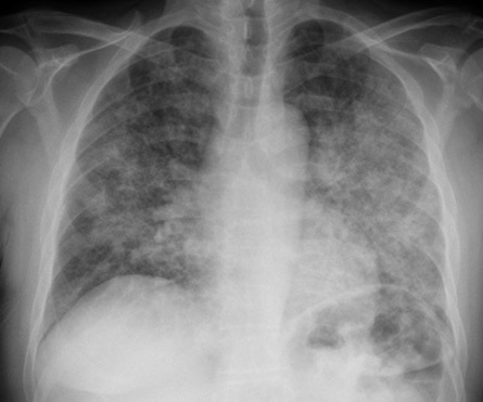
- CXR/CT chest: Imaging may show diffuse, bilateral interstitial infiltrates or a "ground-glass" appearance, which can be misinterpreted as pulmonary oedema. Pneumatoceles and cystic changes can also be seen.
- Elevated lactate dehydrogenase (LDH): LDH levels are often elevated (typically >500 U/L), serving as a non-specific marker of lung injury and inflammation in PCP.
- ABG: Arterial blood gas analysis often shows hypoxaemia (Type 1 respiratory failure). PaO₂ ≤ 9.3 kPa (70 mmHg) on room air indicates severe disease.
- Beta-D-glucan test: This serum marker can be elevated in cases of PCP, though it is not specific and may be elevated in other fungal infections.
Differentials
- Bacterial or viral pneumonia (e.g., influenza, COVID-19).
- Pulmonary embolism if imaging is normal but hypoxia persists.
- Asthma (though wheeze is minimal in PCP).
- Pulmonary oedema, with potential overlapping radiological features.
- HIV-related tuberculosis or mycobacterial infections.
Management
- Stabilize the patient with high-flow oxygen, monitor closely, and initiate HIV testing. Urgent referral to an HIV specialist is essential.
- PCP Prevention: Prophylaxis with Co-trimoxazole (trimethoprim-sulfamethoxazole) 960 mg daily or alternate dosing for those with CD4 < 200 cells/mm3 or those who have previously had PCP.
- PCP Treatment - Mild to Moderate:
- Co-trimoxazole PO 1920 mg TDS (or 90 mg/kg/day in 3 divided doses, rounded to the nearest 480 mg) for 21 days.
- Atovaquone 750 mg BD with food for 21 days if Co-trimoxazole is not tolerated, particularly effective when taken with high-fat meals due to poor bioavailability.
- Dapsone 100 mg PO OD + Trimethoprim 20 mg/kg/day in 3 divided doses for patients unable to tolerate Co-trimoxazole.
- PCP Treatment - Severe:
- Co-trimoxazole IV infusion at 120 mg/kg/day for 3 days, then 90 mg/kg/day for 18 days, divided into 3-4 doses. Transition to oral when appropriate.
- Clindamycin 600 mg IV QDS or 900 mg TDS + Primaquine PO 30 mg OD for patients intolerant to Co-trimoxazole.
- Pentamidine 4 mg/kg IV OD for at least 14 days as a third-line option.
- If PaO₂ ≤ 9.3 kPa or O₂ saturations < 92% on room air, start corticosteroids to reduce inflammation and improve outcomes:
- Prednisolone 40 mg BD for 5 days, then taper to 40 mg OD for 5 days, followed by 20 mg OD for 11 days.
- IV methylprednisolone 40 mg QDS can be used if oral therapy is not feasible.
- Monitor for complications like pneumothorax, which has a poor prognosis in severe cases. Perform CXR if deterioration occurs.
- Ventilatory support may be required due to progressive respiratory failure. High risk of pneumothorax must be managed with careful monitoring.
- Initiate HAART for HIV patients to improve their CD4 count above 200 cells/mm3, reducing recurrence risk and improving long-term survival.
Prognosis and Complications
- Prognosis is better with early diagnosis and treatment. Mortality in untreated cases is high, especially in those with severe hypoxaemia.
- Relapse is possible, particularly in patients who remain severely immunocompromised or have suboptimal adherence to prophylaxis or ART.
- Pneumothorax occurs in 5-10% of severe cases and is associated with a high mortality rate.
- Secondary infections, including bacterial and mycobacterial infections, are common in patients with HIV.
Prevention and Long-Term Management
- Secondary prophylaxis with Co-trimoxazole should continue until the CD4 count is consistently > 200 cells/mm3 for at least 3-6 months on effective ART.
- Educate patients on adherence to ART and regular follow-up to monitor immune status and response to treatment.
- Regular monitoring of lung function and imaging may be required in those with persistent symptoms or complications.
References