ECG - Hyperkalaemia ❤️
Related Subjects:
|ECG Basics
|ECG Axis
|ECG Analysis
|ECG LAD
|ECG RAD
|ECG Low voltage
|ECG Pathological Q waves
|ECG ST/T wave changes
|ECG LBBB
|ECG RBBB
|ECG short PR
|ECG Heart Block
|ECG Asystole and P wave asystole
|ECG QRS complex
|ECG ST segment
|ECG: QT interval
|ECG: LVH
|ECG RVH
|ECG: Bundle branch blocks
|ECG Dominant R wave in V1
|ECG Acute Coronary Syndrome
|ECG Crib sheets
|ECG - LVH
|ECG - STEMI
|ECG Analysis
⚡ Overview
- Hyperkalaemia = Elevated potassium levels in the blood (K⁺ > 5.5 mmol/L).
It can cause profound cardiac conduction abnormalities and life-threatening arrhythmias if untreated. 🚨
📉 ECG Features of Hyperkalaemia
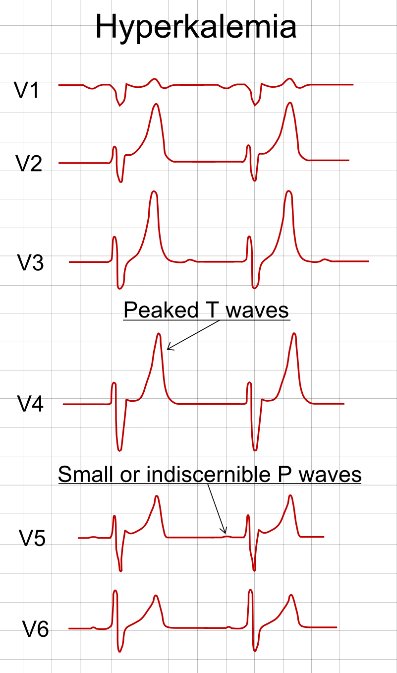
- 🔺 Peaked T Waves: Earliest sign – tall, narrow, "tented" T waves.
- ❌ Absent P Waves: Loss of atrial activity as potassium rises.
- 📏 Broad QRS Complex: Progressive widening; red flag for impending arrest.
- 〰️ Sine Wave Pattern: Fusion of QRS and T ➝ pre-terminal sign.
- 🐢 Bradycardia: Slowed rate due to impaired conduction.
- 💔 Agonal Rhythm ➝ Asystole: Final rhythm before cardiac standstill.
- ⚠️ Cardiac Arrest: High risk of VF/VT, PEA, or asystole.
📊 ECG Progression (Potassium Levels)
| Serum K⁺ (mmol/L) | ECG Change |
|---|
| 5.5 – 6.5 | Peaked T waves, shortened QT interval |
| 6.5 – 7.5 | P wave flattening, PR prolongation |
| 7.5 – 8.5 | QRS widening, sine-wave begins |
| > 8.5 | Ventricular fibrillation, asystole |
📸 ECG Example
Hyperkalaemia on ECG: Tall peaked T waves with broad QRS complexes.
⚡ Emergency Management of Hyperkalaemia
- 1️⃣ Stabilise Myocardium
- IV Calcium Gluconate 10% (10 mL over 2–5 min).
- Notes: Acts within minutes; protects the heart but does not lower serum K⁺.
- 2️⃣ Shift K⁺ Into Cells
- IV Insulin (10 units Actrapid + 50 mL 50% dextrose).
- Nebulised Salbutamol 10–20 mg.
- IV Sodium Bicarbonate (if metabolic acidosis present).
- Notes: Onset within 15–30 min; effect is temporary.
- 3️⃣ Remove K⁺ From Body
- Loop diuretics (if volume replete).
- Potassium binder per local protocol (e.g. sodium zirconium cyclosilicate); calcium resonium is no longer routinely recommended (UKKA 2026).
- Haemodialysis → definitive if refractory or renal failure.
- Notes: Provides permanent removal of potassium.
- 4️⃣ Monitor
- Continuous ECG monitoring.
- Repeat K⁺ every 1–2 hours.
- Look for recurrent rises or ongoing causes.
🔍 Key Clinical Pearls
- Always treat the ECG, not the number ➝ a patient with ECG changes and K⁺ 6.0 mmol/L is more urgent than one with K⁺ 7.0 mmol/L and a normal ECG.
- Check and stop contributing drugs (e.g., ACE inhibitors, ARBs, spironolactone). 💊
- Common in renal failure, acidosis, tissue breakdown (e.g., rhabdomyolysis, tumour lysis). 🧪
