Makindo Medical Notes"One small step for man, one large step for Makindo" |
|
|---|---|
| Download all this content in the Apps now Android App and Apple iPhone/Pad App | |
| MEDICAL DISCLAIMER: The contents are under continuing development and improvements and despite all efforts may contain errors of omission or fact. This is not to be used for the assessment, diagnosis, or management of patients. It should not be regarded as medical advice by healthcare workers or laypeople. It is for educational purposes only. Please adhere to your local protocols. Use the BNF for drug information. If you are unwell please seek urgent healthcare advice. If you do not accept this then please do not use the website. Makindo Ltd. |
Cardiac Embryology
-
| About | Anaesthetics and Critical Care | Anatomy | Biochemistry | Cardiology | Clinical Cases | CompSci | Crib | Dermatology | Differentials | Drugs | ENT | Electrocardiogram | Embryology | Emergency Medicine | Endocrinology | Ethics | Foundation Doctors | Gastroenterology | General Information | General Practice | Genetics | Geriatric Medicine | Guidelines | Haematology | Hepatology | Immunology | Infectious Diseases | Infographic | Investigations | Lists | Microbiology | Miscellaneous | Nephrology | Neuroanatomy | Neurology | Nutrition | OSCE | Obstetrics Gynaecology | Oncology | Ophthalmology | Oral Medicine and Dentistry | Paediatrics | Palliative | Pathology | Pharmacology | Physiology | Procedures | Psychiatry | Radiology | Respiratory | Resuscitation | Rheumatology | Statistics and Research | Stroke | Surgery | Toxicology | Trauma and Orthopaedics | Twitter | Urology
Related Subjects: |Cardiac Anatomy and Physiology |Coronary Artery Anatomy and Physiology |Cardiac Electrophysiology |Cardiac Embryology |Brain Embryology
Cardiac embryology refers to the development of the heart during the early stages of human development. The process is complex and involves the formation and transformation of various structures that will ultimately become the fully functional adult heart.
Key Stages of Heart Development

- Formation of the Cardiogenic Field :
- Occurs in the third week of gestation.
- Mesodermal cells migrate to form the primary heart field, which is located in the splanchnic mesoderm.
- This field will give rise to the heart tube.
- Formation of the Heart Tube :
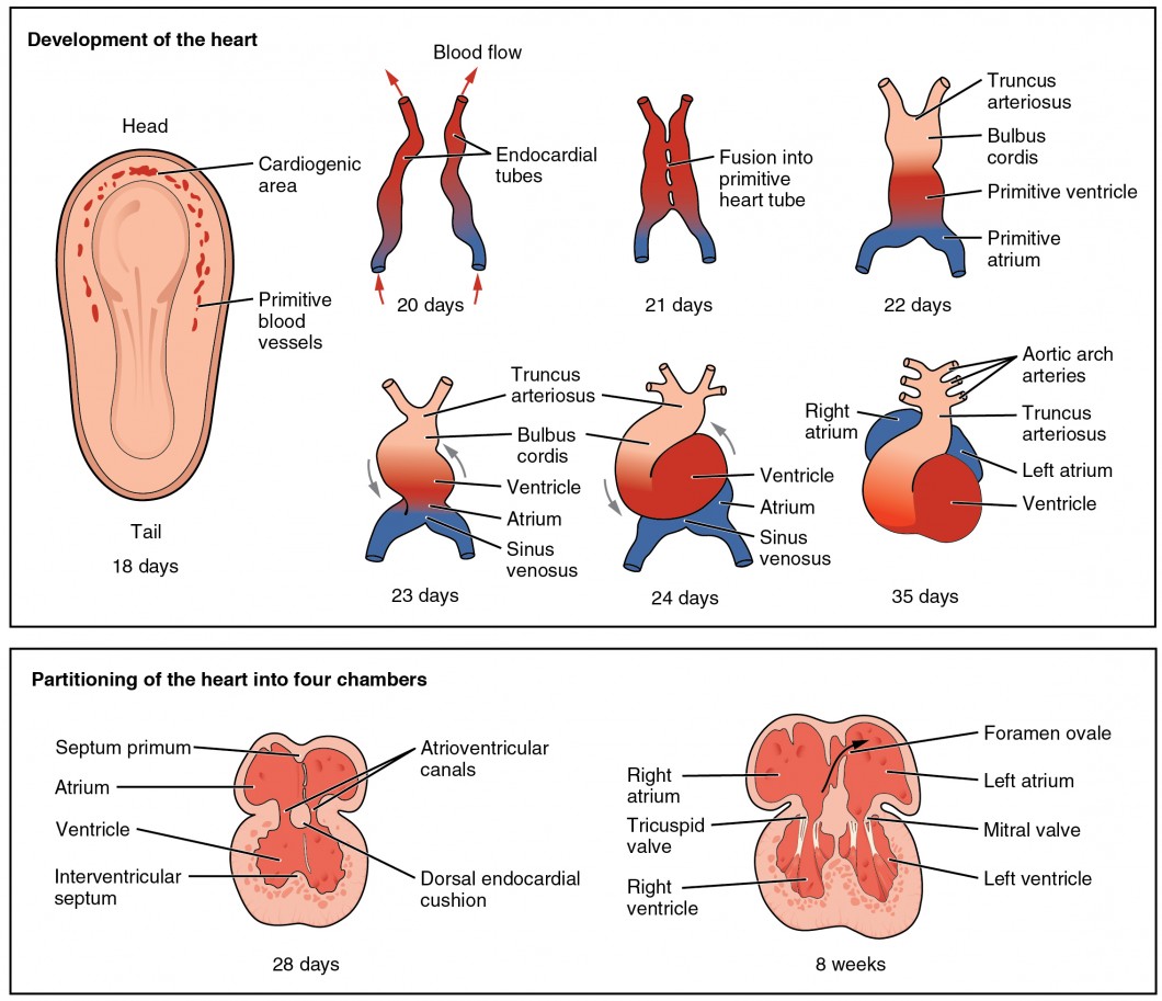
- During the third to fourth week, the bilateral heart fields fuse to form a single heart tube.
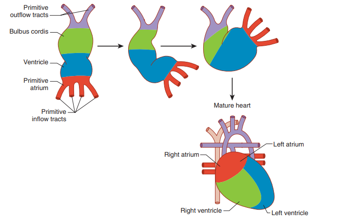
- The heart tube consists of the following regions from head to tail: truncus arteriosus, bulbus cordis, primitive ventricle, primitive atrium, and sinus venosus.
- Looping of the Heart Tube :
- Occurs during the fourth week of development.
- The heart tube undergoes rightward looping, forming an S-shaped structure, which establishes the basic layout of the future heart.
- This process is crucial for proper alignment of the atria and ventricles.
- Septation :
- Septation of the heart occurs between the fourth and eighth weeks.
- Atrial Septation :
- The septum primum grows toward the endocardial cushions, creating the foramen primum.
- The foramen secundum forms before the foramen primum closes, ensuring continuous blood flow between atria.
- The septum secundum forms to the right of the septum primum, creating the foramen ovale.
- Ventricular Septation :
- The muscular interventricular septum grows upward from the floor of the primitive ventricle.
- The membranous interventricular septum forms from the endocardial cushions and conotruncal ridges, completing the separation between the left and right ventricles.
- Outflow Tract Septation :
- The truncus arteriosus is divided into the aorta and pulmonary trunk by the spiral aorticopulmonary septum.
- This septation ensures proper alignment of the aorta with the left ventricle and the pulmonary trunk with the right ventricle.
- Atrial Septation :
- Septation of the heart occurs between the fourth and eighth weeks.
- Formation of the Cardiac Valves :
- Valvulogenesis occurs between the fifth and ninth weeks.
- Atrioventricular valves (mitral and tricuspid) form from the endocardial cushions.
- Semilunar valves (aortic and pulmonary) form from the conotruncal swellings and surrounding mesenchyme.
- Valvulogenesis occurs between the fifth and ninth weeks.
Key Structures in Cardiac Development
- Endocardial Cushions :
- Mesenchymal structures that contribute to the formation of the atrial and ventricular septa, as well as the atrioventricular valves.
- Conotruncal Ridges :
- Form from neural crest cells and contribute to the septation of the outflow tract into the aorta and pulmonary trunk.

Clinical Relevance
- Congenital Heart Defects :
- Abnormalities in cardiac development can lead to congenital heart defects (CHDs), which are the most common type of birth defect.
- Examples include:
- Atrial Septal Defect (ASD) : An opening in the atrial septum, allowing blood flow between the atria.
- Ventricular Septal Defect (VSD) : An opening in the ventricular septum, leading to blood flow between the ventricles.
- Tetralogy of Fallot : A complex defect involving VSD, pulmonary stenosis, right ventricular hypertrophy, and an overriding aorta.
- Transposition of the Great Arteries : The aorta arises from the right ventricle and the pulmonary trunk from the left ventricle, resulting in parallel rather than sequential circulation.
- Genetic Factors :
- Genetic mutations and chromosomal abnormalities can influence heart development, leading to congenital heart defects.
- Conditions such as Down syndrome and DiGeorge syndrome are associated with a higher incidence of CHDs.
- Environmental Factors :
- Maternal factors such as diabetes, infections (e.g., rubella), and exposure to certain medications or toxins can affect cardiac development.
Summary
Cardiac embryology involves a complex sequence of events that transform a simple tube of cells into a fully functional heart. Understanding the stages of heart development and the structures involved is crucial for recognizing and addressing congenital heart defects. Both genetic and environmental factors can influence cardiac development, highlighting the importance of comprehensive prenatal care and early diagnosis.