Makindo Medical Notes"One small step for man, one large step for Makindo" |
|
|---|---|
| Download all this content in the Apps now Android App and Apple iPhone/Pad App | |
| MEDICAL DISCLAIMER: The contents are under continuing development and improvements and despite all efforts may contain errors of omission or fact. This is not to be used for the assessment, diagnosis, or management of patients. It should not be regarded as medical advice by healthcare workers or laypeople. It is for educational purposes only. Please adhere to your local protocols. Use the BNF for drug information. If you are unwell please seek urgent healthcare advice. If you do not accept this then please do not use the website. Makindo Ltd. |
Assessing Coma/Consciousness
-
| About | Anaesthetics and Critical Care | Anatomy | Biochemistry | Cardiology | Clinical Cases | CompSci | Crib | Dermatology | Differentials | Drugs | ENT | Electrocardiogram | Embryology | Emergency Medicine | Endocrinology | Ethics | Foundation Doctors | Gastroenterology | General Information | General Practice | Genetics | Geriatric Medicine | Guidelines | Haematology | Hepatology | Immunology | Infectious Diseases | Infographic | Investigations | Lists | Microbiology | Miscellaneous | Nephrology | Neuroanatomy | Neurology | Nutrition | OSCE | Obstetrics Gynaecology | Oncology | Ophthalmology | Oral Medicine and Dentistry | Paediatrics | Palliative | Pathology | Pharmacology | Physiology | Procedures | Psychiatry | Radiology | Respiratory | Resuscitation | Rheumatology | Statistics and Research | Stroke | Surgery | Toxicology | Trauma and Orthopaedics | Twitter | Urology
Related Subjects: |Assessing Coma and Management |Glasgow Coma scale |Acute Poisoning |Trauma: Traumatic Brain Head Injury (TBI) |Acute Anaphylaxis |Basic Life Support |Advanced Life Support |Acute Stroke Assessment |Brain Herniation syndromes |Haemorrhagic stroke |Acutely ill patient |Distributive Shock |Hypovolaemic or Haemorrhagic Shock |Obstructive Shock |Septic Shock and Sepsis |Shock (General Assessment)
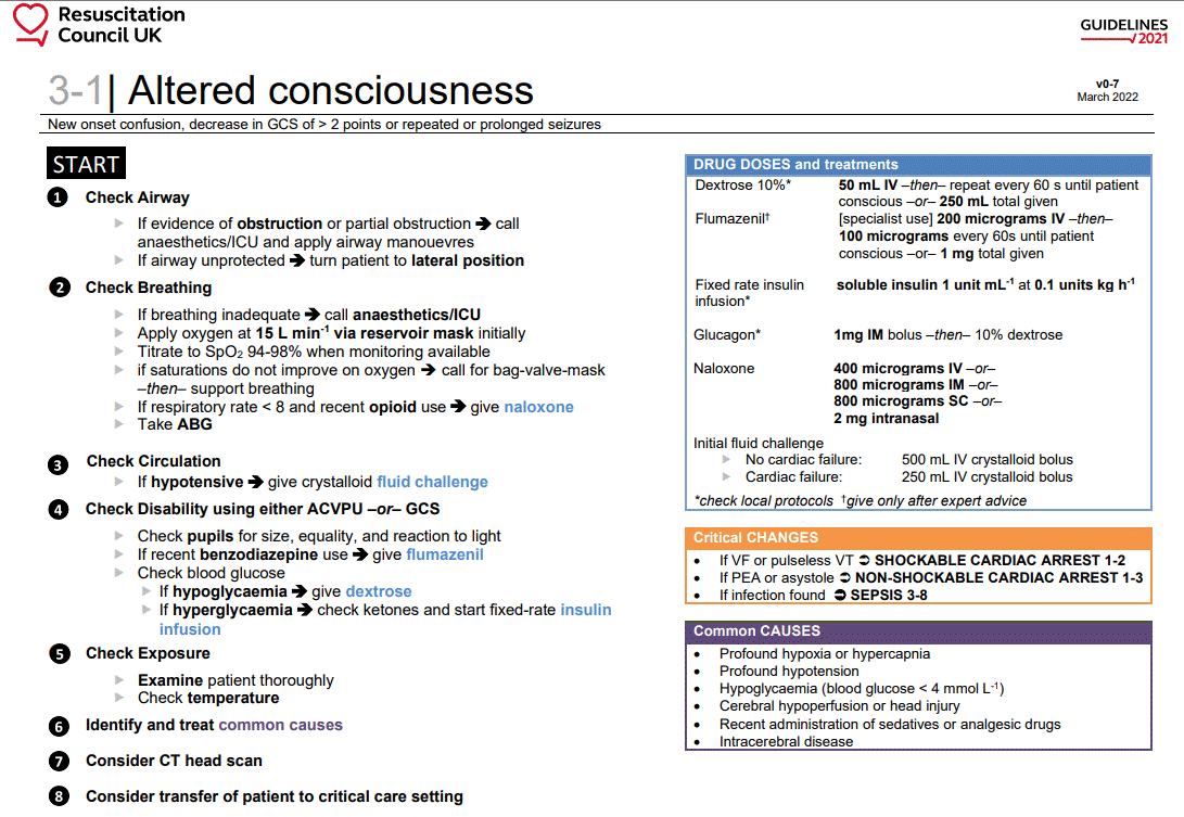
⚠️ If the cause of coma is uncertain (e.g. CT negative), always consider giving the CAND bundle:
💉 Cefotaxime (meningitis cover),
💊 Aciclovir (HSV encephalitis),
💉 Naloxone (opiates),
🍬 Dextrose ± Pabrinex (thiamine, prevent Wernicke’s).
This covers the major reversible causes until further information is available.
| 🛠️ Coma Management Overview |
|---|
|
🛌 Recovery Position


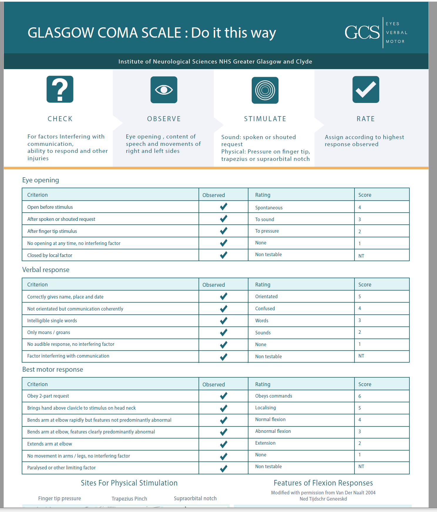
📏 Assessment of Coma: GCS & Pupils


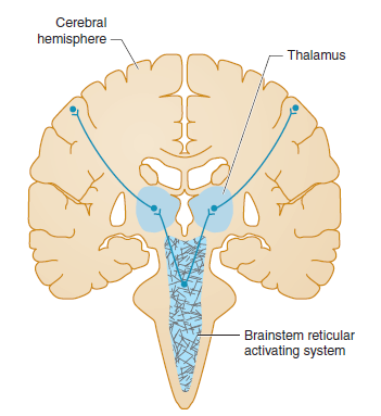
🧠 Pathophysiology of Coma (ARAS)
- Consciousness requires intact Ascending Reticular Activating System (ARAS) + cortex.
- ARAS = brainstem nuclei → thalamus → cortex.
- Coma occurs with ↑ ICP, bilateral cortical/thalamic damage, or brainstem dysfunction.
- Pupils give localising clues: 🔴 Bilateral dilated, fixed → herniation/brainstem failure. 🔵 Pinpoint → pons or opioids. ⚖️ Unequal → structural lesion compressing CN III.

🧩 Consciousness depends on ARAS (midbrain/pons) + both cerebral hemispheres. Structural lesions or diffuse metabolic insults can both produce coma.
🧪 Differential Diagnosis: CT & Glucose First
| Cause | Clues | Management |
|---|---|---|
| 🍬 Hypoglycaemia | Low CBG | IV Dextrose |
| 💊 Opiate toxicity | Pinpoint pupils | IV Naloxone |
| 🧠 Stroke | Focal neurology | CT head, stroke pathway |
| ⚡ Post-ictal | History of seizure | Recovery position, treat status |
| 🦠 Meningitis/Encephalitis | Fever, neck stiffness | Cefotaxime + Aciclovir |
| ☠️ CO poisoning | Exposure, headache, cherry-red skin | 100% O₂ ± hyperbaric |
| 🤕 Head injury | History of trauma | CT head/C-spine |
| 💧 Hyponatraemia | Na <115, seizures | Hypertonic saline |
| 🧬 Sepsis | Fever, shock | IV Tazocin + Gentamicin |
| 📟 NCSE | Unresponsive, subtle eye movements | EEG, Lorazepam |
| 💥 SDH/EDH | Head trauma | Neurosurgery + Mannitol |
| 🦟 Cerebral malaria | Travel history | IV Quinine |
⚡ Pupils in Coma
| Pupil Type | Description | Causes |
|---|---|---|
| Normal | 3–4 mm, reactive | Metabolic causes |
| Thalamic | ~2 mm, reactive | Thalamic compression |
| Fixed dilated | >7 mm, unreactive | Herniation, CN III compression |
| Midsized fixed | ~5 mm, unreactive | Midbrain lesion |
| Pinpoint | 1–1.5 mm, minimal reaction | Pons, opioids, organophosphates |
| Asymmetric | Anisocoria | Structural lesion, CN III palsy |
🚨 Signs of Trauma
- 🐼 Raccoon eyes (periorbital bruising).
- 🎧 Battle’s sign (mastoid bruising).
- 🩸 Haemotympanum.
- 💧 CSF rhinorrhoea/otorrhoea (confirm with β-2 transferrin).
- 👆 Palpable depressed skull fracture.
📊 Pathophysiologic Patterns
| Feature | Supratentorial | Subtentorial | Diffuse/Metabolic |
|---|---|---|---|
| Pupils | Normal or dilated with herniation | Midsized (midbrain), pinpoint (pons) | Normal/reactive; pinpoint (opiates); dilated (anticholinergics) |
| Eye movements | May show gaze preference | Abnormal adduction/abduction | Usually preserved unless sedated |
| Motor | Often asymmetric | Asymmetric (uni) or symmetric (bi) | Usually symmetric |
📚 Teaching Commentary
Coma is a syndrome, not a diagnosis. Always think structural vs metabolic. 🔍 Pupils are the fastest localising sign: normal = metabolic, abnormal = structural. 🇬🇧 In UK practice, juniors should remember: “CAND for coma if CT is normal” → Cefotaxime, Aciclovir, Naloxone, Dextrose + Pabrinex. This covers the major reversible killers until more is known. GCS < 8 = intubate. Always immobilise the neck if trauma suspected.
References
Cases — Coma (Reduced GCS)
- Case 1 — Hypoglycaemia: A 58-year-old man with type 2 diabetes on insulin is found unresponsive at home. Capillary glucose is 1.8 mmol/L. GCS 6/15. Pupils equal and reactive. After IV 20% dextrose, consciousness improves rapidly. Teaching point: Always check glucose in any unconscious patient (“D” in ABCDE). Hypoglycaemia is a reversible cause of coma, with rapid recovery if treated promptly. 🍬
- Case 2 — Traumatic brain injury: A 22-year-old motorcyclist is brought in after a high-speed collision. He has unequal pupils (left fixed and dilated), GCS 5/15, and irregular breathing. CT shows an acute left extradural haematoma with midline shift. Teaching point: Low GCS after trauma should prompt urgent neuroimaging. Extradural haematomas may have a classic “lucid interval,” but once coma develops, urgent neurosurgical decompression is required. 🧠
- Case 3 — Sepsis with encephalopathy: A 75-year-old woman with community-acquired pneumonia becomes progressively drowsy. On arrival: temp 39.5°C, BP 80/50, HR 120, GCS 7/15. She localises to pain but does not obey commands. Teaching point: Sepsis-associated encephalopathy is common in older adults; reduced GCS in this setting signals severe illness and high mortality risk. Urgent ABC resuscitation, antibiotics, and source control are priorities. 🌡️