
| Download the amazing global Makindo app: ✅ Means NICE/National Guidelines 2026 compliant Android | Apple | |
|---|---|
| MEDICAL DISCLAIMER: Educational use only. Not for diagnosis or management. See below for full disclaimer. |
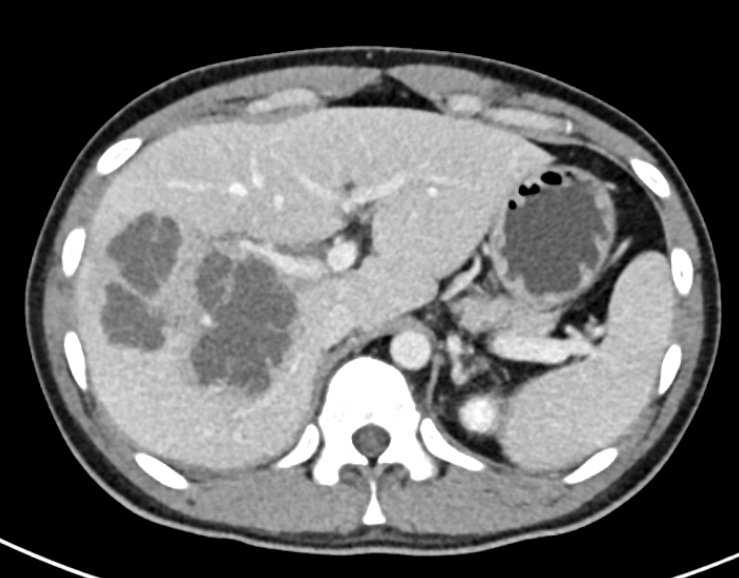
Liver abscess ✅
Related Subjects: |Chronic liver disease |Cirrhosis |Alkaline phosphatase (ALP) |Liver Function Tests |Ascites Assessment and Management |Budd-Chiari syndrome |Autoimmune Hepatitis |Primary Biliary Cirrhosis |Primary Sclerosing Cholangitis |Wilson disease |Hereditary Haemochromatosis |Alpha-1 Antitrypsin (AAT) deficiency |Non alcoholic steatohepatitis (NASH) |Spontaneous Bacterial Peritonitis |Alcoholism and Alcoholic Liver Disease |Hepatitis C
🩺 Liver Abscess = a pus-filled cavity in the liver parenchyma. ⚡ Pathophysiology: hepatocyte injury → local inflammation → neutrophilic infiltration → liquefaction → abscess formation. Remember: Klebsiella pneumoniae abscesses may disseminate to the eye causing endophthalmitis 👁️.
📖 About
- Pus-filled lesion(s) within liver tissue; usually right lobe due to portal venous drainage patterns.
- ⚠️ Differentials: metastases, simple cysts, hepatocellular carcinoma.
🦠 Aetiology
- 🧫 Pyogenic (bacterial) – polymicrobial: E. coli, Klebsiella, Streptococcus, Staphylococcus; often secondary to cholangitis, cholecystitis, diverticulitis, appendicitis, trauma.
- 🪱 Amoebic – Entamoeba histolytica; more common in endemic areas.
- 🍄 Fungal – Candida spp.; immunocompromised patients.
- 🐕 Echinococcus – hydatid cysts; risk of anaphylaxis on rupture.
📌 Causes, Features, Diagnosis & Treatment
| Cause | Clinical Features | Diagnosis | Initial Treatment (UK, NICE-aligned) |
|---|---|---|---|
| Pyogenic | Fever, rigors, RUQ pain ± shoulder tip pain, jaundice, hepatomegaly, malaise, weight loss | USS/CT, blood cultures, aspiration & microbiology | IV broad-spectrum antibiotics (adjust per culture), image-guided drainage; surgery if refractory |
| Amoebic | Gradual fever, RUQ pain, ± diarrhoea, weight loss | USS (often single right lobe), serology (anti-amoebic IgG), aspiration if diagnosis unclear | Metronidazole → luminal agent (e.g., diloxanide furoate); drain only if large/persistent |
| Fungal | Fever, RUQ pain, weight loss; immunocompromised | CT (multiple abscesses), blood culture, biopsy | Systemic antifungals (amphotericin B, fluconazole); drainage if large |
| Echinococcus | Often asymptomatic; cyst rupture → anaphylaxis, urticaria, shock | USS/CT, serology (ELISA, WB), PCR if difficult | Surgical resection if feasible; otherwise PAIR (puncture, aspiration, injection ethanol, re-aspiration) + albendazole |
🧑⚕️ Clinical Features
- 🌡️ Fever, chills, sweats, malaise
- 🤢 Anorexia, weight loss
- RUQ tenderness, hepatomegaly
- Right shoulder or pleuritic chest pain, hiccups
- ⚠️ Hydatid cyst rupture → urticaria, angioedema, anaphylaxis
🔍 Differentials
- Hepatocellular carcinoma or liver metastases
- Simple liver cysts, polycystic liver disease
⚠️ Complications
- Sepsis ± septic shock
- Empyema/peritonitis if rupture
- Endophthalmitis (especially Klebsiella)
🧪 Investigations
- 📊 Bloods: ↑ WCC, ↑ ALP (95%), ± ↑ AST/ALT, low albumin
- 🧾 Coagulation: PT/INR
- 🩸 Blood cultures (aerobic & anaerobic)
- 🖼 Imaging: USS/CT (right lobe common)
- 🪱 Entamoeba: stool microscopy (low yield), serology (EIA/PCR)
- 🐕 Echinococcus: serology (ELISA/WB), PCR if needed
💊 Management (UK, NICE-aligned)
- 🛡️ Stabilisation: ABC, IV fluids, analgesia
- 🧫 IV broad-spectrum antibiotics → adjust per culture & sensitivities
- 🩺 Image-guided drainage (needle/catheter)
- 🪱 Amoebic abscess: Metronidazole → luminal agent (diloxanide)
- 🍄 Fungal: Amphotericin B / azole antifungals; drainage if large
- 🐕 Echinococcus: surgical resection if feasible; otherwise PAIR + albendazole
💡 Teaching tip: Always consider source of infection (biliary, portal, systemic), assess for immunosuppression, and monitor for sepsis. Early imaging and culture-guided therapy improve outcomes. Referral to hepatology or infectious disease recommended for complex cases.
| The content on this website is provided for educational and informational purposes only to support exam preparation (e.g., MLA, MRCP, USMLE) and learning. This is NOT medical advice, diagnosis, treatment, or professional guidance. It does not replace consultation with a qualified healthcare professional, official guidelines (e.g., NICE, GMC, BNF), or supervised clinical practice. Always verify information with current, authoritative sources. Makindo and its contributors accept no liability for any reliance on this content, including errors, omissions, or any resulting harm, loss, or consequences. By using this site, you agree to these terms. |
|
|
Categories
- About
- Acute Medicine
- Anaesthetics and Critical Care
- Anatomy
- Anatomy and Physiology
- Biochemistry
- Book
- Cardiology
- Collections
- CompSci
- Crib Sheets
- Critical care
- Dental
- Dermatology
- Differentials
- Drugs
- ENT
- Electrocardiogram
- Embryology
- Emergency Medicine
- Endocrinology
- Ethics
- Foundation Doctors
- GCSE
- Gastroenterology
- General Practice
- Genetics
- Geriatric Medicine
- Geriatrics
- Guidelines
- Haematology
- Hepatology
- Immunology
- Infectious Diseases
- Infographic
- Investigations
- Lists
- MRCP
- Mandatory Training
- Medical Students
- Microbiology
- Nephrology
- Neurology
- Neurosurgery
- Nutrition
- OSCE
- Obstetrics Gynaecology
- Oncology
- Ophthalmology
- Oral Medicine and Dentistry
- Orthopaedics
- Paediatrics
- Palliative
- Palliative Care
- Pathology
- Pharmacology
- Physiology
- Procedures
- Psychiatry
- Public Health
- Radiology
- Respiratory
- Resuscitation
- Revision Topics
- Rheumatology
- Statistics and Research
- Stroke
- Surgery
- Toxicology
- Trauma and Orthopaedics
- USMLE
- Urology
- Vascular Surgery