
🦴 Bone scintigraphy (bone scan) is a nuclear medicine test that detects abnormal bone metabolism. ⚡ It is different from a DEXA scan, which measures bone density rather than turnover.
🔎 About
- Uses technetium-99m-labelled bisphosphonate (99mTc) as a tracer, which accumulates in areas of high bone turnover.
- Imaged by a gamma camera, producing whole-body bone maps highlighting pathological activity.
- Highly sensitive but not specific - abnormal uptake can occur in infection, tumour, trauma, or inflammation.
⚙️ How a Bone Scan Works
- IV injection of 99mTc-bisphosphonate.
- Tracer binds to bone mineral at sites of increased osteoblastic activity.
- Gamma camera detects radiation → produces images of metabolic “hot spots.”
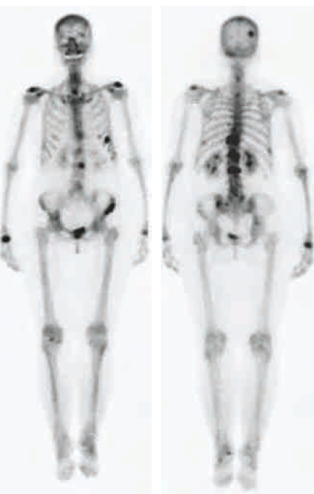
Example: Breast cancer with multiple bone metastases showing widespread tracer uptake.
📸 Imaging Phases
- Immediate (Perfusion Phase): Seconds after injection – reflects blood flow. Increased uptake in infection, inflammation, or tumour vascularity.
- Delayed (Remodelling Phase): 2–4 hours later – reflects osteoblastic activity and bone turnover. Main diagnostic phase.
🧾 Diagnostic Applications
- Skeletal metastases: 🦠 Sensitive for prostate (osteoblastic) and breast cancer spread.
- Primary bone tumours / Benign disease: Osteosarcoma, Paget’s disease, osteoarthritis.
- Stress fractures & Osteomalacia: Identifies Looser’s zones and early stress injuries.
- Complex Regional Pain Syndrome (CRPS): Early metabolic changes detectable before X-ray changes.
- Sclerosing bone disorders: e.g. hypertrophic pulmonary osteoarthropathy (HPOA).
- Spondyloarthritides: Uptake at sacroiliac joints and entheses (insertion points).
📊 Comparison: Bone Scan vs DEXA vs MRI
| Test | Main Purpose | Strengths | Limitations |
|---|---|---|---|
| Bone Scintigraphy (99mTc) | Detects ↑ bone turnover / metastases | Whole-body survey, highly sensitive | Low specificity (tumour vs infection vs trauma may look similar) |
| DEXA | Measures bone density (osteoporosis) | Gold standard for fracture risk assessment | No info on bone metabolism |
| MRI | Soft tissue, marrow pathology | Excellent detail, no radiation | Localised, expensive, less suited for whole-body survey |
💡 Clinical Pearls
- 🔥 Hot spots: areas of ↑ uptake (tumour, infection, fracture, inflammation).
- ❄️ Cold spots: areas of ↓ uptake (e.g., avascular necrosis, myeloma lesions).
- Always interpret alongside history, X-ray, CT, or MRI for specificity.