Makindo Medical Notes"One small step for man, one large step for Makindo" |
|
|---|---|
| Download all this content in the Apps now Android App and Apple iPhone/Pad App | |
| MEDICAL DISCLAIMER: The contents are under continuing development and improvements and despite all efforts may contain errors of omission or fact. This is not to be used for the assessment, diagnosis, or management of patients. It should not be regarded as medical advice by healthcare workers or laypeople. It is for educational purposes only. Please adhere to your local protocols. Use the BNF for drug information. If you are unwell please seek urgent healthcare advice. If you do not accept this then please do not use the website. Makindo Ltd. |
Granulomas
-
| About | Anaesthetics and Critical Care | Anatomy | Biochemistry | Cardiology | Clinical Cases | CompSci | Crib | Dermatology | Differentials | Drugs | ENT | Electrocardiogram | Embryology | Emergency Medicine | Endocrinology | Ethics | Foundation Doctors | Gastroenterology | General Information | General Practice | Genetics | Geriatric Medicine | Guidelines | Haematology | Hepatology | Immunology | Infectious Diseases | Infographic | Investigations | Lists | Microbiology | Miscellaneous | Nephrology | Neuroanatomy | Neurology | Nutrition | OSCE | Obstetrics Gynaecology | Oncology | Ophthalmology | Oral Medicine and Dentistry | Paediatrics | Palliative | Pathology | Pharmacology | Physiology | Procedures | Psychiatry | Radiology | Respiratory | Resuscitation | Rheumatology | Statistics and Research | Stroke | Surgery | Toxicology | Trauma and Orthopaedics | Twitter | Urology
Related Subjects: | Chronic Granulomatous Disease | Granulomatous Diseases
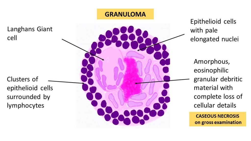
🧩 Granulomatous diseases are conditions in which the immune system forms granulomas — organised clusters of macrophages, lymphocytes, and giant cells — as a defence mechanism when unable to eliminate a pathogen or trigger. ⚠️ Granulomas are a non-specific pathological pattern and must be interpreted in clinical context.

🔬 Characteristics of Granulomas
- Structure 🧱 :
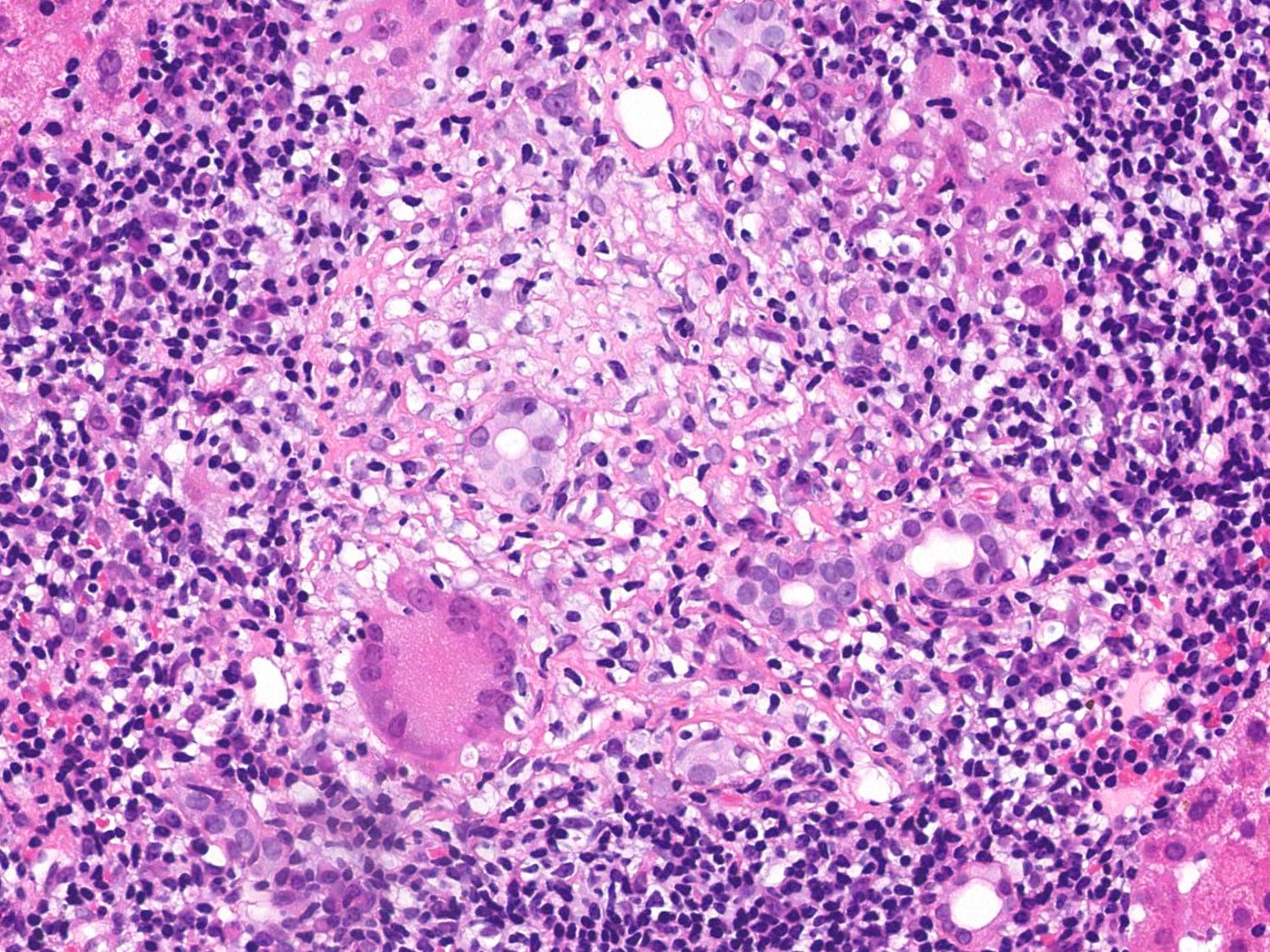
- Aggregates of macrophages (epithelioid cells), lymphocytes, and multinucleated giant cells.
- Caseating granulomas (central necrosis, cheese-like appearance) → classic of TB & fungal infections.
- Non-caseating granulomas (no necrosis) → sarcoidosis, Crohn’s disease.
- Occasional calcification in chronic granulomas.
- Formation 🔄 :
- Triggered by persistent antigenic stimulus.
- Macrophages → epithelioid cells → multinucleated giant cells (Langhans type or foreign-body type).
- T-helper (CD4+) cells, TNF-α, and interferon-γ play central roles.

🧾 Common Granulomatous Diseases
- Tuberculosis 🫁 :
- Mycobacterium tuberculosis → caseating granulomas.
- Symptoms: chronic cough, fever, night sweats, weight loss.
- May cavitate and spread (miliary TB).
- Sarcoidosis 🌿 :
- Autoimmune → non-caseating granulomas.
- Affects lungs, lymph nodes, eyes, skin.
- Symptoms: cough, dyspnoea, erythema nodosum, uveitis.
- Granulomatosis with Polyangiitis (GPA) 🩸 :
- Necrotising vasculitis with granulomas.
- Affects ENT, lungs, kidneys.
- Symptoms: sinusitis, haematuria, cough, pulmonary nodules.
- Lab: c-ANCA (PR3 antibodies).
- Leprosy 🧕 :
- Mycobacterium leprae → granulomas in skin, nerves, airway.
- Symptoms: anaesthetic patches, neuropathy, deformities.
- Histoplasmosis 🍂 :
- Fungal → caseating granulomas in lung & lymph nodes.
- Can mimic TB; disseminates in immunocompromised hosts.
- Cryptococcosis 🍄 :
- Cryptococcus neoformans → lung + brain granulomas.
- Symptoms: cough, chest pain, meningitis in HIV.
- Cat-Scratch Disease 🐱 :
- Bartonella henselae → granulomatous lymphadenitis.
- Symptoms: fever, fatigue, tender lymph nodes after cat scratch/bite.
🔍 Diagnosis
- Clinical 🩺 : Symptoms vary by organ system.
- Imaging 🖼️ : CXR/CT (lung granulomas), MRI/CT (CNS involvement).
- Lab 🧪 :
- Blood tests (inflammatory markers, serology).
- Tuberculin skin test / interferon-gamma release assay (TB).
- ANCA (vasculitis), ACE level (sarcoidosis).
- Histology 🔬 : Biopsy shows granuloma type (caseating vs non-caseating).
💊 Treatment
- Infectious 🌡️ :
- TB → multi-drug therapy (RIPE regimen).
- Leprosy → rifampicin + dapsone ± clofazimine.
- Fungal infections → antifungals (Itraconazole, Amphotericin B).
- Autoimmune 🛡️ :
- Sarcoidosis/GPA → corticosteroids, methotrexate, azathioprine, biologics (rituximab for GPA).
- Supportive 🩹 :
- Pain control, oxygen therapy for lung disease, immunosuppression monitoring.
⚠️ Clinical Pearls
- Always distinguish infectious vs autoimmune → treatment differs radically (antibiotics vs immunosuppression).
- Caseating granulomas → usually TB/fungal. Non-caseating → sarcoid, Crohn’s, GPA.
- Some granulomas calcify → appear as incidental CXR/CT findings.
- Chronic granulomas can lead to fibrosis and organ dysfunction.
📚 Summary
Granulomatous diseases represent a spectrum of infectious 🦠, autoimmune 🌿, and inflammatory 🔥 conditions. Histology (caseating vs non-caseating), microbiology, and systemic context guide diagnosis. Treatment may involve antimicrobials, antifungals, corticosteroids, or immunosuppressants, tailored to the underlying cause.