Makindo Medical Notes"One small step for man, one large step for Makindo" |
|
|---|---|
| Download all this content in the Apps now Android App and Apple iPhone/Pad App | |
| MEDICAL DISCLAIMER: The contents are under continuing development and improvements and despite all efforts may contain errors of omission or fact. This is not to be used for the assessment, diagnosis, or management of patients. It should not be regarded as medical advice by healthcare workers or laypeople. It is for educational purposes only. Please adhere to your local protocols. Use the BNF for drug information. If you are unwell please seek urgent healthcare advice. If you do not accept this then please do not use the website. Makindo Ltd. |
Acutely Ill Patient
-
| About | Anaesthetics and Critical Care | Anatomy | Biochemistry | Cardiology | Clinical Cases | CompSci | Crib | Dermatology | Differentials | Drugs | ENT | Electrocardiogram | Embryology | Emergency Medicine | Endocrinology | Ethics | Foundation Doctors | Gastroenterology | General Information | General Practice | Genetics | Geriatric Medicine | Guidelines | Haematology | Hepatology | Immunology | Infectious Diseases | Infographic | Investigations | Lists | Microbiology | Miscellaneous | Nephrology | Neuroanatomy | Neurology | Nutrition | OSCE | Obstetrics Gynaecology | Oncology | Ophthalmology | Oral Medicine and Dentistry | Paediatrics | Palliative | Pathology | Pharmacology | Physiology | Procedures | Psychiatry | Radiology | Respiratory | Resuscitation | Rheumatology | Statistics and Research | Stroke | Surgery | Toxicology | Trauma and Orthopaedics | Twitter | Urology
Related Subjects: |Assessing Chest Pain |Acute Coronary Syndrome (ACS) General |Aortic Dissection |Pulmonary Embolism |Acute Pericarditis |Diffuse Oesophageal Spasm |Gastro oesophageal reflux |Oesophageal Perforation Rupture |Pericardial Effusion Tamponade |Pneumothorax |Tension Pneumothorax |Lactic acidosis
🚨 Profoundly sick patient: Call for help early! Stay calm 😌, focus on ABCs, and work systematically — you can stabilise most situations if you think step by step.
| ⚡ Acutely Ill Patient – First Priorities |
|---|
|
📖 Introduction
- ⏱ Work quickly & logically — always think ABC.
- 🔍 POCUS is invaluable (pneumothorax, tamponade, LVF, AAA).
- 🧃 Start O₂, gain IV access, attach monitoring early.
- ✋ Always check a central pulse & monitor rhythm.
- 💉 Decompress suspected tension pneumothorax immediately.
- 🩸 Major haemorrhage → activate massive transfusion protocol.
- 🧪 High-yield tests (if safe): ECG, CXR, ABG, Lactate, Bedside Echo, CT guided by presentation.
- 💊 Give IV antibiotics early if sepsis suspected (e.g. Co-Amoxiclav 1.2 g IV qds ± Gentamicin, per local policy).
- 🚽 Catheterise shocked patients to monitor urine output.
❓ If patient not improving – always ask
- 🤔 Is the diagnosis correct?
- 🧩 Could there be >1 diagnosis?
- 📊 Do I need more info/tests urgently?
- ☎️ Should I escalate now — registrar/consultant/ICU?
🫁 Airway
- 🚨 Suspected epiglottitis/stridor → do not examine airway. Fast bleep Anaesthetics/ENT. Give high-flow O₂.
- 😮💨 Simple obstruction → Head tilt, chin lift, remove debris/foreign body.
- 💊 Opiates → Naloxone 0.4 mg IV (repeat; max 10 mg).
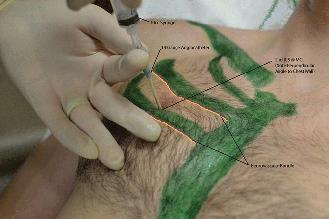
- 🫁 Tension Pneumothorax → Needle decompression 2nd ICS mid-clavicular line.
- 💓 Acute LVF → Crackles, tachypnoea → GTN + IV Furosemide. ECG & CXR. Urgent reperfusion if STEMI.
- 🫀 Acute PE → Chest pain, hypoxia, shock → Bedside Echo/CTPA. Heparin ± thrombolysis if haemodynamically unstable.
- 😤 Acute Severe Asthma → tiring, hypercapnia → urgent ITU review.
- 🌪️ COPD Exacerbation → Controlled O₂ (24–28%), consider NIV if Type 2 RF.
- 🦠 Pneumonia/COVID → Hypoxia + CXR changes.
- 😱 Panic attack → diagnosis of exclusion. Low PaCO₂.
💨 Decompressing Tension Pneumothorax

🩸 Hypotension / Shock
- 💧 No clear cause → Trial 500 ml IV N-Saline (250 ml if breathless/pulmonary oedema).
- 🩸 Haemorrhagic shock → GI bleed, varices → urgent blood + endoscopy/surgical input.
- 🦠 Septic shock → High lactate + infection → start Sepsis 6, IV antibiotics <30 mins.
- 💥 Leaking AAA → Sudden abdo/back pain, pulsatile mass → vascular surgery.
- 👩🍼 Ectopic pregnancy → Shock + abdo pain in fertile female → urgent gynae/surgery.
- 🫀 Cardiogenic shock → Post-MI or myocarditis. Echo, ECG. Consider inotropes + PCI.
- 🧩 Tamponade → Raised JVP, muffled HS, hypotension → urgent pericardiocentesis.
- ⚡ Anaphylaxis → IM Adrenaline 500 mcg + IV fluids.
- 🩻 Dissection → Tearing pain, BP asymmetry → CT Aorta, BP control, cardiothoracics.
- ☣️ Meningococcal septicaemia → Purpura, sepsis → IV Ceftriaxone + ITU.
- 🌑 Addisonian crisis → Shock, hyperpigmentation, hyponatraemia → IV Hydrocortisone.
❤️ Chest Pain
- 🫀 ACS → ECG, troponin, O₂, morphine, aspirin, PCI if STEMI.
- 🩻 Dissection → CT Aortogram, avoid anticoagulation until excluded.
- 🫁 PE → CTPA ± Echo if unstable.
- 🪠 Oesophageal rupture/Boerhaave → Severe pain post-vomit → surgical emergency.
🧠 Comatose
- 🍬 Hypoglycaemia → 50 ml 50% dextrose IV.
- 💊 Opiates → Naloxone up to 10 mg.
- 🧠 SOL/Stroke → CT Head ± LP. Consider aciclovir/ceftriaxone empirically if encephalitis/meningitis possible.
- 🍷 Alcohol/Drug intoxication → Supportive, antidotes if available.
- ⚡ Post-ictal or NCSE → EEG, benzodiazepines if seizing.
- 🌍 Cerebral malaria → travel history crucial.
⚡ Arrhythmias
- 📉 Bradycardia (<50 bpm + hypotension) → Atropine → pacing if no response.
- 📈 Tachycardia (>120 bpm + shock) → DC Cardioversion.
- 📊 Always get ECG and follow ALS algorithm.
🤕 Abdominal Pain Emergencies
- 💥 Leaking AAA → Shock + abdo pain → vascular surgery.
- 🔥 Pancreatitis → Severe pain, Grey-Turner’s/Cullen’s signs.
- 🕳️ Perforated viscus → Free air under diaphragm, rigid abdomen → urgent surgery.
- 👩🍼 Ectopic pregnancy → Shocked fertile female → Gynae emergency.
- 🩸 GI bleed → Endoscopy, PPI, correct coagulopathy, blood as required.
Cases — Acutely Ill Patient Assessment (ABCDE)
- Case 1 — Sepsis from Pneumonia 🌡️: A 67-year-old man presents with confusion, fever 39.2°C, RR 32, BP 85/50, O₂ sats 86% on air. Exam: crackles over right lung base. Assessment: - A: patent, - B: tachypnoea, hypoxia, - C: hypotension, tachycardia, - D: GCS 13, - E: febrile, no rash. Management: High-flow O₂, IV fluids, IV antibiotics within 1 hr, blood cultures, lactate, escalate to critical care if unstable.
- Case 2 — Acute Severe Asthma 😮💨: A 23-year-old woman presents with severe breathlessness, unable to complete sentences. RR 36, O₂ sats 88%, widespread wheeze, PEFR 25% predicted. BP 110/70, HR 128. Assessment: - A: speaking in short phrases, - B: severe bronchospasm, poor air entry, - C: tachycardic but perfusing, - D: alert, - E: no other findings. Management: High-flow O₂, nebulised salbutamol + ipratropium, IV hydrocortisone, IV magnesium, prepare for intubation if tiring.
- Case 3 — Acute Pulmonary Oedema 💔: A 72-year-old woman with known heart failure presents with acute dyspnoea, orthopnoea, frothy pink sputum. O₂ sats 82%, BP 170/100, widespread crackles. Assessment: - A: patent, - B: severe hypoxia, pulmonary oedema, - C: hypertension, tachycardia, - D: anxious but alert, - E: peripheral oedema. Management: Sit upright, high-flow O₂, IV furosemide, IV nitrates if BP allows, morphine if distressed, consider CPAP.
- Case 4 — Anaphylaxis 🐝: A 34-year-old man stung by a wasp develops rapid swelling of lips, wheeze, stridor, BP 70/40, O₂ sats 84%. Assessment: - A: threatened (stridor), - B: bronchospasm + hypoxia, - C: shock, - D: anxious but oriented, - E: urticarial rash. Management: IM adrenaline 0.5 mg, high-flow O₂, IV fluids, IV hydrocortisone + chlorphenamine, prepare for airway management.
- Case 5 — Hypoglycaemia in Diabetic Patient 🍬: A 55-year-old man with type 1 diabetes found drowsy at home. GCS 8, HR 110, BP 110/70. Capillary glucose 1.9 mmol/L. Assessment: - A: patent, - B: normal, - C: tachycardic but perfusing, - D: reduced GCS, - E: pale, sweaty. Management: Immediate IV glucose 10–20% (or IM glucagon if no IV access), monitor response, identify cause (missed meal, insulin overdose, infection).
- Case 6 — Septic Shock from Urosepsis 🌡️: A 75-year-old woman presents with fever, rigors, and confusion. BP 80/40, HR 130, SpO₂ 92% RA. Exam: suprapubic tenderness, oliguria. Assessment: - A: patent, - B: tachypnoea 28/min, SpO₂ 92%, - C: hypotensive, tachycardic, mottled, - D: GCS 12, - E: febrile 39°C. Management: Sepsis 6 (O₂, cultures, IV antibiotics, IV fluids, lactate, urine output); urgent critical care input.
- Case 7 — Upper GI Bleed 🩸: A 58-year-old man with alcohol excess collapses with haematemesis and melaena. BP 85/50, HR 140, pale and clammy. Assessment: - A: patent, - B: tachypnoea, SpO₂ 95%, - C: shocked (tachycardia, hypotension), - D: GCS 14, - E: melaena + haematemesis. Management: Large-bore IV access ×2, bloods (group & save, crossmatch), IV fluids, transfusion as needed, IV PPI, urgent endoscopy once stabilised.
- Case 8 — Tension Pneumothorax ⚡: A 32-year-old man with sudden pleuritic chest pain and dyspnoea after trauma. Exam: tachycardia 150, tracheal deviation, absent breath sounds on right, hyperresonant chest. Assessment: - A: patent, - B: severe distress, absent right breath sounds, - C: hypotension 75/40, JVP raised, - D: anxious, - E: chest trauma. Management: Immediate needle decompression (2nd intercostal space, midclavicular line) → chest drain insertion; O₂; monitor for recurrence.
- Case 9 — Acute Stroke 🧠: A 70-year-old woman presents with sudden left-sided weakness and slurred speech. BP 180/100, HR 88, SpO₂ 95% RA. GCS 14. Assessment: - A: patent, - B: normal, - C: hypertensive, regular, - D: focal neurology (left arm/leg weakness, dysarthria), - E: no trauma, normothermic. Management: FAST + stroke call; urgent CT head; thrombolysis if within 4.5h and no contraindications; aspirin 300 mg once haemorrhage excluded.
- Case 10 — Hyperkalaemia with ECG Changes ⚡: A 66-year-old man on haemodialysis misses 2 sessions, presents with weakness. ECG: tall peaked T waves, widened QRS. BP 100/60, HR 60. Assessment: - A: patent, - B: normal, - C: bradycardic, borderline perfusion, - D: GCS 15, - E: no sepsis, no bleed. Management: IV calcium gluconate, IV insulin + dextrose, nebulised salbutamol, consider calcium resonium or dialysis. Continuous cardiac monitoring.
Teaching Commentary 🧠
Assessing the acutely ill patient requires a structured ABCDE approach. - A: airway patency (threatened? stridor?). - B: respiratory distress, hypoxia, RR. - C: perfusion (pulse, BP, JVP, cap refill). - D: GCS, pupils, glucose. - E: temperature, rashes, exposure. Early recognition and intervention save lives. Always reassess after each step and call for senior/critical care help early in deteriorating patients.