Subclavian Vein Thrombosis (SCVT)
Related Subjects:
| Pulmonary Embolism
| Deep Vein Thrombosis
| DVT/PE in Pregnancy
| CTPA
🩺 The subclavian vein lies beneath the clavicle and continues from the axillary vein, merging with the internal jugular vein to form the brachiocephalic vein. ⚠️ Thrombosis here (SCVT) is an important form of upper-extremity DVT.
📖 About
- 🩸 Subclavian vein thrombosis (SCVT) = upper-limb DVT.
- 🔗 Causes: trauma, catheters, malignancy, or inherited clotting disorders.
🧬 Aetiology (Virchow’s Triad)
- 🩸 Hypercoagulability (blood composition changes).
- 🌀 Stasis/turbulent flow.
- 🧱 Endothelial injury/damage to the vessel wall.
📌 Causes
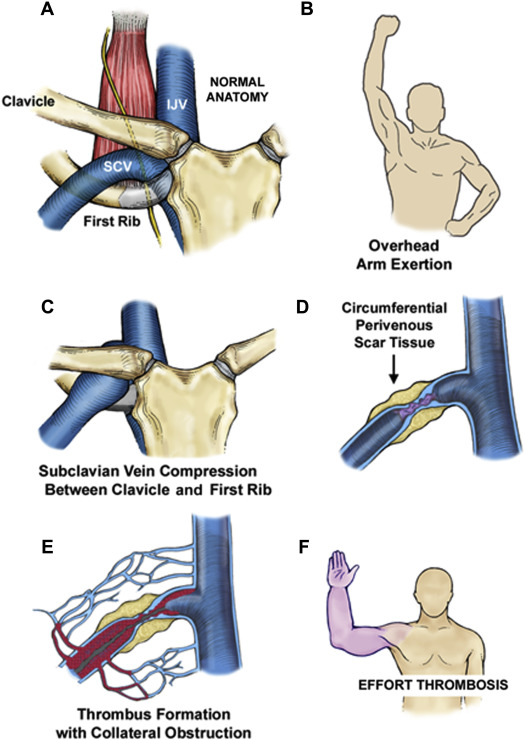
- Primary SCVT 🏋️♂️ (Effort-Induced):
- 💪 Paget–Schroetter Syndrome: Young, healthy, after vigorous activity.
- ❓ Idiopathic: sometimes due to hidden malignancy.
- Secondary SCVT 🏥:
- 🦴 Trauma (clavicle fracture, compression).
- 💉 Central venous catheters, pacemakers, dialysis lines, TPN.
- 🛏️ Prone positioning during surgery.
- 🧬 Inherited thrombophilia (e.g., Factor V Leiden, Protein C/S deficiency).
- 🎗️ Lung cancer (esp. Pancoast tumours).
🩺 Clinical Features
- 💪 Arm swelling, oedema, heaviness.
- 🌐 Dilated veins (neck, shoulder, arm).
- 🔵 Cyanosis in fingers/hand.
- 📈 Possible lymphadenopathy (cervical/axillary).
- 👩⚕️ Breast exam if malignancy suspected.
🧪 Investigations
- 🧾 Bloods: FBC, U&E, ESR/CRP, ↑ D-dimer.
- 🧬 Thrombophilia screen (esp. young pts).
- 🖼️ Imaging: CXR, CT/MRI venography.
- 🔊 Duplex ultrasound = first-line.
- 💉 Contrast venography = gold standard.
- 🎗️ Mammography if breast cancer suspected.
⚠️ Complications
- 🫁 Pulmonary embolism (PE).
- 🦵 Post-thrombotic syndrome (chronic swelling, stasis ulcers).
- 🦠 Septic thrombophlebitis.
- 🫀 Superior vena cava (SVC) syndrome.
- 🚫 Loss of central venous access (catheter occlusion).
🔍 Differential Diagnoses
- 🫀 Superior vena cava syndrome.
- 🧬 Lymphatic obstruction.
- 🔥 Cellulitis.
- 🪓 Thoracic outlet obstruction.
- 🎗️ Pancoast tumour.
- ☠️ Necrotizing fasciitis.
- 🩸 Superficial thrombophlebitis.
🛠️ Management (Cause-Dependent)
- 💉 Anticoagulation: Start LMWH → warfarin/DOACs.
- 🧼 Thrombolysis: Catheter-directed in effort-induced/catheter-related cases.
- 🪡 Stenting: For significant venous narrowing.
- 🕵️ Investigate: Underlying malignancy or thrombophilia.
- ❌ Remove catheter: If device is the culprit (after acute phase).
📚 References
🧑⚕️ Case Examples - Subclavian Vein Thrombosis (SCVT)
-
Case 1 (Effort-induced / Paget-Schroetter syndrome): 🏋️
A 22-year-old competitive rower presents with sudden swelling, heaviness, and cyanosis of the right arm after intense training. Dilated superficial chest wall veins are visible.
Analysis: “Effort thrombosis” of the subclavian vein due to repetitive overhead arm activity causing venous compression.
Diagnosis: Duplex ultrasound confirms acute SCVT.
Management: Anticoagulation, catheter-directed thrombolysis if severe, and later surgical decompression (first rib resection) to prevent recurrence.
-
Case 2 (Catheter-related SCVT): 💉
A 65-year-old man with a central venous catheter for chemotherapy develops left arm swelling, pain, and erythema over 48 hours.
Analysis: Indwelling catheters are the commonest cause of secondary SCVT, provoking local thrombosis.
Diagnosis: Duplex/CT venography shows thrombus around catheter in the left subclavian vein.
Management: Anticoagulation (LMWH/DOAC), remove catheter if no longer essential, oncology team review.
-
Case 3 (Malignancy-associated SCVT): 🎗️
A 58-year-old woman with known breast cancer presents with right arm swelling, facial plethora, and dilated chest wall veins.
Analysis: Cancer is a strong pro-thrombotic state; extension towards the SVC suggests SVC obstruction.
Diagnosis: CT venography shows right subclavian vein thrombosis with proximal extension.
Management: Anticoagulation, oncology input, consider stenting if symptomatic SVC obstruction.
