Makindo Medical Notes"One small step for man, one large step for Makindo" |
|
|---|---|
| Download all this content in the Apps now Android App and Apple iPhone/Pad App | |
| MEDICAL DISCLAIMER: The contents are under continuing development and improvements and despite all efforts may contain errors of omission or fact. This is not to be used for the assessment, diagnosis, or management of patients. It should not be regarded as medical advice by healthcare workers or laypeople. It is for educational purposes only. Please adhere to your local protocols. Use the BNF for drug information. If you are unwell please seek urgent healthcare advice. If you do not accept this then please do not use the website. Makindo Ltd. |
Optic Nerve (Cranial Nerve II) and tract
-
| About | Anaesthetics and Critical Care | Anatomy | Biochemistry | Cardiology | Clinical Cases | CompSci | Crib | Dermatology | Differentials | Drugs | ENT | Electrocardiogram | Embryology | Emergency Medicine | Endocrinology | Ethics | Foundation Doctors | Gastroenterology | General Information | General Practice | Genetics | Geriatric Medicine | Guidelines | Haematology | Hepatology | Immunology | Infectious Diseases | Infographic | Investigations | Lists | Microbiology | Miscellaneous | Nephrology | Neuroanatomy | Neurology | Nutrition | OSCE | Obstetrics Gynaecology | Oncology | Ophthalmology | Oral Medicine and Dentistry | Paediatrics | Palliative | Pathology | Pharmacology | Physiology | Procedures | Psychiatry | Radiology | Respiratory | Resuscitation | Rheumatology | Statistics and Research | Stroke | Surgery | Toxicology | Trauma and Orthopaedics | Twitter | Urology
Related Subjects: |Olfactory Nerve |Optic Nerve |Oculomotor Nerve |Trochlear Nerve |Trigeminal Nerve |Abducent Nerve |Facial Nerve |Vestibulocochlear Nerve |Glossopharyngeal Nerve |Vagus Nerve |Accessory Nerve |Hypoglossal Nerve
The optic nerve, also known as cranial nerve II, is responsible for transmitting visual information from the retina to the brain. It is a vital part of the visual system, enabling the sense of sight.
Anatomy of the Optic Nerve
- Origin :
- The optic nerve originates from the ganglion cells in the retina of the eye.
- Course :
- The optic nerve exits the orbit through the optic canal.
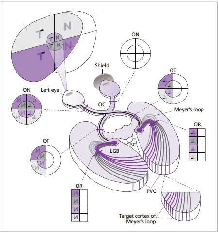
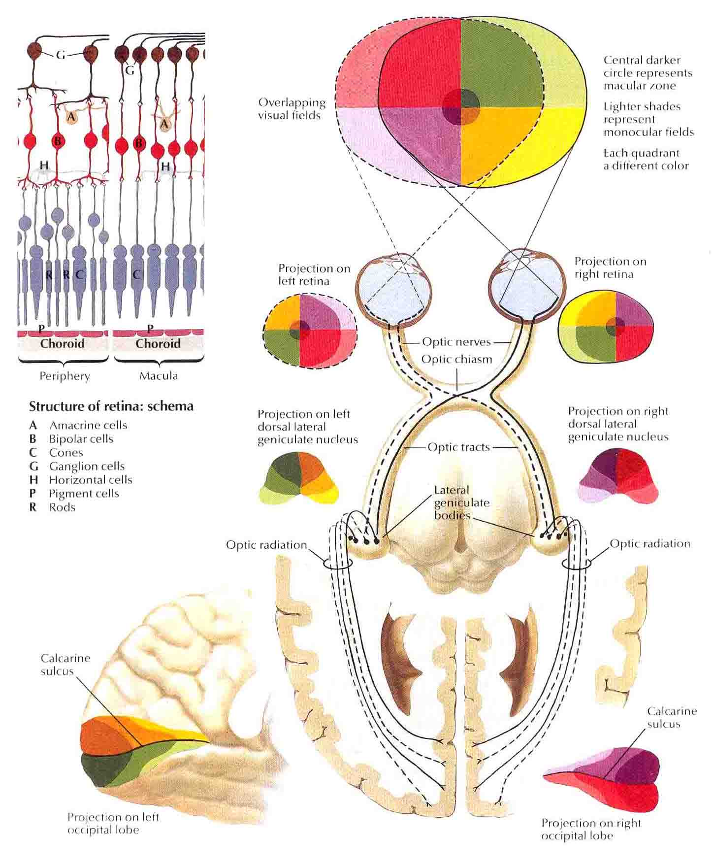
- It then converges with the optic nerve from the opposite eye at the optic chiasm, located at the base of the brain.
- At the optic chiasm, nerve fibers partially decussate (cross over) to the opposite side.
- Post-chiasmal fibers continue as the optic tracts, which terminate in the lateral geniculate nucleus (LGN) of the thalamus.

Functions of the Optic Nerve
- Visual Information Transmission :
- The optic nerve transmits visual information from the photoreceptors in the retina to the brain for processing.
- This information includes aspects such as light intensity, colour, and spatial resolution.
- Visual Reflexes :
- Involved in reflexive responses such as the pupillary light reflex, where the pupil constricts in response to bright light.
Clinical Relevance
- Optic Neuritis :
- Inflammation of the optic nerve, often associated with multiple sclerosis.
- Symptoms include sudden vision loss, pain with eye movement, and changes in colour perception.
- Treatment may include corticosteroids to reduce inflammation.
- Glaucoma :
- A group of eye conditions that damage the optic nerve, often due to increased intraocular pressure.
- Can lead to gradual vision loss and, if untreated, blindness.
- Treatment includes medications, laser therapy, or surgery to lower intraocular pressure.
- Optic Atrophy :
- Degeneration of the optic nerve fibers, leading to vision loss.
- Caused by various conditions, including ischaemia, trauma, or neurodegenerative diseases.
- Visual Field Defects :
- Damage to the optic nerve or its pathways can result in specific patterns of visual field loss.
- Bitemporal hemianopia is a classic defect resulting from a lesion at the optic chiasm, often caused by pituitary tumours.
- Evaluation and Diagnosis :
- Ophthalmologic examination, including visual acuity testing and fundoscopic examination, to assess the optic nerve head.
- Imaging studies such as MRI or CT scans to evaluate the optic nerve and brain structures.
- Visual field testing to identify areas of vision loss.
- Optical coherence tomography (OCT) to assess the thickness of the retinal nerve fiber layer.
Summary
The optic nerve (cranial nerve II) is crucial for vision, transmitting visual information from the retina to the brain. It originates from the retinal ganglion cells, exits the orbit through the optic canal, and forms the optic chiasm where fibers partially cross. The optic nerve is involved in visual information processing and reflexes. Clinical conditions affecting the optic nerve include optic neuritis, glaucoma, and optic atrophy, which can lead to vision loss. Thorough evaluation and timely treatment are essential for managing optic nerve-related disorders.
Overview of Visual Field Defects
Visual field defects are areas of diminished or absent vision within the normal visual field. They can result from various conditions affecting the eye, optic nerve, or brain. Understanding these defects is crucial for diagnosing and managing underlying conditions.
Types of Visual Field Defects
- Central Scotoma :
- Description: Loss of vision in the center of the visual field.
- Common Causes: Macular degeneration, optic neuritis, multiple sclerosis.
- Peripheral Field Loss :
- Description: Loss of vision in the periphery of the visual field.
- Common Causes: Glaucoma, retinitis pigmentosa.
- Bitemporal Hemianopia :
- Description: Loss of vision in the outer (temporal) halves of both visual fields.
- Common Causes: Lesion at the optic chiasm, often caused by pituitary tumours.
- Homonymous Hemianopia :
- Description: Loss of vision in the same half of the visual field in both eyes (right or left).
- Common Causes: Lesions in the contralateral temporal, parietal or occipital cortex (e.g., stroke, brain tumours).
- Quadrantanopia :
- Description: Loss of vision in a quarter of the visual field.
- Common Causes: Lesions in the contralateral temporal, parietal or occipital cortex (e.g., stroke, brain tumours). Temporal - superior defect, parietal and inferior defect
- Altitudinal Field Defect :
- Description: Loss of vision in the upper or lower half of the visual field.
- Common Causes: Anterior ischaemic optic neuropathy (AION), retinal artery occlusion.
Causes of Visual Field Defects
- Optic Nerve Disorders :
- Optic neuritis
- Glaucoma
- Optic atrophy
- Ischaemic optic neuropathy
- Retinal Disorders :
- Retinal detachment
- Macular degeneration
- Retinitis pigmentosa
- Retinal vein or artery occlusion
- Lesions at the Optic Chiasm :
- Pituitary tumours
- Craniopharyngiomas
- Meningiomas
- Post-Chiasmal Lesions :
- Stroke
- Brain tumours
- Trauma
- Multiple sclerosis
- Other Causes :
- Migraines
- Hypertension
- Diabetes
Evaluation and Diagnosis
- Clinical History :
- Detailed patient history including onset, duration, and characteristics of vision loss.
- Associated symptoms such as headache, pain, or neurological deficits.
- Past medical history including conditions like diabetes, hypertension, or multiple sclerosis.
- Ophthalmologic Examination :
- Visual acuity testing
- Fundoscopic examination
- Visual field testing using perimetry
- Imaging Studies :
- MRI or CT scans to evaluate the optic nerve, chiasm, and brain structures
- Optical coherence tomography (OCT) to assess the retinal nerve fiber layer
- Electrophysiological Tests :
- Visual evoked potentials (VEP) to assess the function of the visual pathways
Management
- Treatment of Underlying Causes :
- Medical or surgical treatment for conditions like glaucoma, retinal detachment, or tumours
- Management of systemic diseases such as diabetes or hypertension
- Vision Rehabilitation :
- Low vision aids and adaptive techniques to help patients cope with vision loss
- Regular Monitoring :
- Follow-up exams to monitor the progression of visual field defects
Summary
Visual field defects are areas of impaired vision that can result from various conditions affecting the eye, optic nerve, or brain. Types of defects include central scotoma, peripheral field loss, bitemporal hemianopia, homonymous hemianopia, quadrantanopia, and altitudinal field defects. Proper evaluation, including clinical history, ophthalmologic examination, imaging, and electrophysiological tests, is essential for diagnosing and managing these defects. Treatment focuses on addressing the underlying cause and providing vision rehabilitation to help patients adapt to their vision loss.
