Pityriasis or Tinea versicolor infections
Related Subjects:
|Nikolsky's sign
|Koebner phenomenon
|Erythema Multiforme
|Pyoderma gangrenosum
|Erythema Nodosum
|Dermatitis Herpetiformis
|Lichen Planus
|Acanthosis Nigricans
|Acne Rosacea
|Acne Vulgaris
|Alopecia
|Vitiligo
|Urticaria
|Basal Cell Carcinoma
|Malignant Melanoma
|Squamous Cell Carcinoma
|Mycosis Fungoides (Sezary Syndrome)
|Xeroderma pigmentosum
|Bullous Pemphigoid
|Pemphigus Vulgaris
|Seborrheic Dermatitis
|Pityriasis/Tinea versicolor infections
|Pityriasis rosea
|Scabies
|Dermatomyositis
|Toxic Epidermal Necrolysis
|Stevens-Johnson Syndrome
|Atopic Eczema/Atopic Dermatitis
|Psoriasis
📖 About
- Pityriasis (Tinea) Versicolor is a common superficial fungal infection caused by yeasts of the Malassezia genus.
- Seen mainly in adolescents and young adults 👩🦱👨🦱, especially in warm and humid climates 🌴.
- Most patients are otherwise healthy, though immunocompromised individuals are at increased risk.
- Previously called Malassezia furfur infection.
🧬 Aetiology & Pathophysiology
- Caused by Pityrosporum orbiculare (round yeast form) and Pityrosporum ovale (oval form).
- Thrives in oily (sebaceous) areas – the organism metabolises skin lipids (free fatty acids & triglycerides).
- Alters melanocyte function → causes hypo- or hyperpigmentation due to uneven melanin production.
- Relapse is common because the organism is part of the normal skin flora.
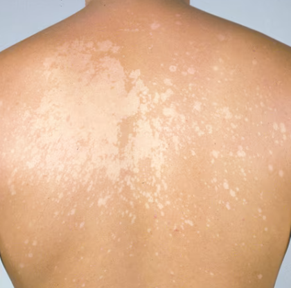
👀 Clinical Features
- Usually asymptomatic, but may cause mild pruritus.
- Multiple small, circular macules – colour may vary (white, pink, brown).
- Often on trunk, neck, upper arms; more obvious after sun exposure ☀️ (hypopigmented patches fail to tan).
- Risk factors: oily skin, sweating, immunosuppression, humid climates.
🔎 Investigations
- Wood’s lamp: yellow-green fluorescence ✨ of affected areas.
- Microscopy (KOH prep): shows the “spaghetti and meatballs” pattern (hyphae + spores).
- Skin biopsy: rarely needed, only if atypical.
💊 Management
- Topical therapy first-line:
- Ketoconazole 2% shampoo – applied scalp → thighs, left 5 min, once daily for 3 days (or single application).
- Alternatives: Selenium sulphide or other azole creams (ketoconazole/clotrimazole BD for small areas).
- Systemic therapy: Reserved for extensive/refractory disease:
- Itraconazole 200 mg daily × 7 days → ~90% cure rate at 4 weeks.
- Griseofulvin ineffective ❌.
- Recurrence is common: intermittent use of medicated shampoo (weekly/monthly) as prophylaxis.
- Note: pigmentary changes may take months to resolve – reassure patients that this does not reflect treatment failure.
✅ Key Exam Pearls
- Caused by Malassezia species → not dermatophytes.
- “Spaghetti and meatballs” appearance on microscopy 🍝.
- Wood’s lamp = yellow-green fluorescence.
- First-line = ketoconazole shampoo, not griseofulvin.
- Pigmentary changes may persist for months despite cure.
