
| Download the amazing global Makindo app: ✅ Means NICE/National Guidelines 2026 compliant Android | Apple | |
|---|---|
| MEDICAL DISCLAIMER: Educational use only. Not for diagnosis or management. See below for full disclaimer. |
Knee Joint Structure and Form
Related Subjects: |Shoulder Joint Structure and Form |Knee Joint Structure and Form |Wrist Joint Structure and Form
The knee is the largest synovial joint in the body and functions as a modified hinge joint. It primarily allows flexion and extension, with small degrees of rotation when flexed. Although superficially simple, it is biomechanically complex, relying heavily on ligaments and surrounding muscles for stability rather than bony congruence.
🦴 Articulating Bones
- Femur – medial and lateral condyles.
- Tibia – tibial plateau (weight-bearing surface).
- Patella – sesamoid bone within quadriceps tendon; improves mechanical advantage of knee extension.
🧵 Menisci
- Medial meniscus – C-shaped, less mobile, attached to MCL (more commonly injured).
- Lateral meniscus – more circular, more mobile.
- Functions:
- Load distribution and shock absorption.
- Joint stability and proprioception.
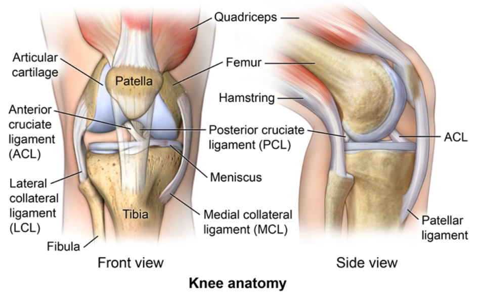
🛡️ Ligaments (Primary Stabilizers)
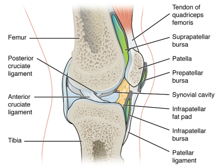
Intracapsular (Cruciate Ligaments)
- ACL – prevents anterior translation of tibia on femur; taut in extension.
- PCL – prevents posterior translation of tibia; stronger than ACL.
Extracapsular
- MCL – resists valgus stress; attached to medial meniscus.
- LCL – resists varus stress; not attached to lateral meniscus.
- Patellar ligament – continuation of quadriceps tendon to tibial tuberosity.
- Oblique popliteal ligament – posterior reinforcement.
- Arcuate ligament complex – posterolateral stability.
💪 Muscles Acting on the Knee
Anterior Compartment (Extension)
- Quadriceps femoris (rectus femoris, vastus medialis, vastus lateralis, vastus intermedius) – primary extensors; femoral nerve.
Posterior Compartment (Flexion)
- Hamstrings (biceps femoris, semitendinosus, semimembranosus) – flex knee; sciatic nerve.
- Gastrocnemius – assists flexion; tibial nerve.
- Popliteus – “unlocks” knee from extension; tibial nerve.
🧵 Important Tendons
- Quadriceps tendon (above patella).
- Patellar tendon (below patella).
- Pes anserinus (sartorius, gracilis, semitendinosus insertion).
- Iliotibial band (lateral stabiliser).
🩸 Blood Supply
- Genicular branches of the popliteal artery form periarticular anastomosis.
- Descending genicular artery (from femoral artery).
⚡ Nerve Supply
- Femoral nerve – anterior capsule.
- Tibial nerve – posterior capsule.
- Common peroneal nerve – lateral aspect.
- Obturator nerve – medial contribution.
🔄 Biomechanics
The knee exhibits a “screw-home” mechanism: during the last 20° of extension, the tibia externally rotates relative to the femur, locking the joint. Popliteus reverses this during flexion. Stability is greatest in full extension due to ligament tension.
⚠️ Clinical Correlations
- ACL tear – pivoting injury; positive Lachman test.
- PCL injury – dashboard injury.
- Meniscal tear – locking/catching sensation.
- MCL injury – valgus stress injury.
- Osteoarthritis – medial c
| The content on this website is provided for educational and informational purposes only to support exam preparation (e.g., MLA, MRCP, USMLE) and learning. This is NOT medical advice, diagnosis, treatment, or professional guidance. It does not replace consultation with a qualified healthcare professional, official guidelines (e.g., NICE, GMC, BNF), or supervised clinical practice. Always verify information with current, authoritative sources. Makindo and its contributors accept no liability for any reliance on this content, including errors, omissions, or any resulting harm, loss, or consequences. By using this site, you agree to these terms. |
|
|
Categories
- About
- Acute Medicine
- Anaesthetics and Critical Care
- Anatomy
- Anatomy and Physiology
- Biochemistry
- Book
- Cardiology
- Collections
- CompSci
- Crib Sheets
- Critical care
- Dental
- Dermatology
- Differentials
- Drugs
- ENT
- Electrocardiogram
- Embryology
- Emergency Medicine
- Endocrinology
- Ethics
- Foundation Doctors
- GCSE
- Gastroenterology
- General Practice
- Genetics
- Geriatric Medicine
- Geriatrics
- Guidelines
- Haematology
- Hepatology
- Immunology
- Infectious Diseases
- Infographic
- Investigations
- Lists
- MRCP
- Mandatory Training
- Medical Students
- Microbiology
- Nephrology
- Neurology
- Neurosurgery
- Nutrition
- OSCE
- Obstetrics Gynaecology
- Oncology
- Ophthalmology
- Oral Medicine and Dentistry
- Orthopaedics
- Paediatrics
- Palliative
- Palliative Care
- Pathology
- Pharmacology
- Physiology
- Procedures
- Psychiatry
- Public Health
- Radiology
- Respiratory
- Resuscitation
- Revision Topics
- Rheumatology
- Statistics and Research
- Stroke
- Surgery
- Toxicology
- Trauma and Orthopaedics
- USMLE
- Urology
- Vascular Surgery