
| Download the amazing global Makindo app: ✅ Means NICE/National Guidelines 2026 compliant Android | Apple | |
|---|---|
| MEDICAL DISCLAIMER: Educational use only. Not for diagnosis or management. See below for full disclaimer. |
Dermatology terms
Related Subjects:
|Nikolsky's sign
|Koebner phenomenon
|Erythema Multiforme
|Pyoderma gangrenosum
|Erythema Nodosum
|Dermatitis Herpetiformis
|Lichen Planus
|Acanthosis Nigricans
|Acne Rosacea
|Acne Vulgaris
|Alopecia
|Vitiligo
|Urticaria
|Basal Cell Carcinoma
|Malignant Melanoma
|Squamous Cell Carcinoma
|Mycosis Fungoides (Sezary Syndrome)
|Xeroderma pigmentosum
|Bullous Pemphigoid
|Pemphigus Vulgaris
|Seborrheic Dermatitis
|Pityriasis/Tinea versicolor infections
|Pityriasis rosea
|Scabies
|Dermatomyositis
|Toxic Epidermal Necrolysis
|Stevens-Johnson Syndrome
|Atopic Eczema/Atopic Dermatitis
|Psoriasis
| Name | Description |
|---|---|
| 🌸 Dermatosis | Any cutaneous lesion or group of lesions. A nonspecific umbrella term for skin disease. |
| 🔥 Dermatitis (Eczema) | Inflammation of the skin. Eczema often used interchangeably with atopic dermatitis. |
| 🤧 Pruritus | Itching of the skin. |
| 🔴 Erythema | Redness due to vascular congestion or increased perfusion. |
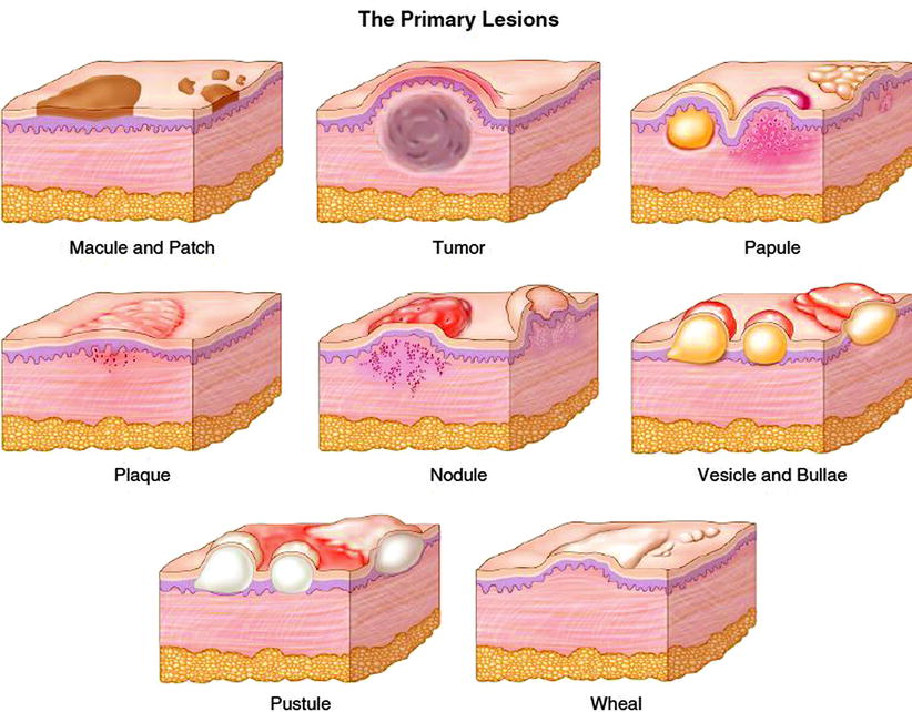
| ⚪ Macule | Flat, circumscribed change in skin colour without elevation/depression.
>2 cm = patch. Examples: Café-au-lait spots, Mongolian spot, freckles. |
| 🔵 Papule | Solid, elevated lesion ≤0.5 cm. May arise from deposits, infiltrates, or cellular hyperplasia.
Papulosquamous lesions = papules with scaling. |
| 🔘 Nodule | Larger, deeper papule, usually >0.5 cm, located in dermis, subcutaneous tissue, or epidermis.
Examples: Xanthoma, metastatic deposits. |
| 🟪 Plaque | Flat-topped, elevated lesion ≥2 cm. Often formed by coalescence of papules. Surface area > height. |
| 💭 Wheal | Transient, raised, edematous lesion (urticarial). Often erythematous and itchy. Size varies from mm → cm. Seen in hives. |
| 💧 Vesicle / Bulla | Clear fluid-filled lesions.
Vesicle: ≤0.5 cm. Bulla: >0.5 cm. Superficial = flaccid. Example: herpes (vesicles), bullous pemphigoid (bullae). |
| 🟡 Pustule | Elevated lesion filled with purulent fluid (white, yellow, or green).
Example: acne pustules. |
| 🩸 Abscess | Localized pus collection within tissue cavity, often with necrosis. |
| 🎈 Cyst | Closed sac containing liquid or semisolid material. Usually resilient on palpation. |
| 📉 Atrophy | Thinning of epidermis/dermis → depressed skin.
Example: striae (“stretch marks”). |
| 🪨 Sclerosis | Diffuse or circumscribed hardening of skin due to dermal/subcutaneous induration. |
| ⬇️ Erosion | Loss of epidermis only. |
| ⚠️ Ulcer | Loss of epidermis + dermis (may extend deeper). If due to scratching → “excoriation”. |
| 〰️ Fissure | Linear cleavage in skin surface (variant of erosion/ulcer).
Example: heel fissures. |
| 🌿 Desquamation | Scaling/exfoliation of the stratum corneum. |
| ➕ Scar | Fibrous tissue after ulceration.
May be hypertrophic, atrophic, or cribriform (pitted). |
| 🩹 Crust (“Scab”) | Dried serum, pus, or blood.
Yellow = serum, green = purulent, brown/red = blood. Hallmark of pyogenic infection. |
| 🪵 Lichenification | Chronic epidermal thickening with exaggerated skin markings, often from scratching/rubbing. |
📚 References
| The content on this website is provided for educational and informational purposes only to support exam preparation (e.g., MLA, MRCP, USMLE) and learning. This is NOT medical advice, diagnosis, treatment, or professional guidance. It does not replace consultation with a qualified healthcare professional, official guidelines (e.g., NICE, GMC, BNF), or supervised clinical practice. Always verify information with current, authoritative sources. Makindo and its contributors accept no liability for any reliance on this content, including errors, omissions, or any resulting harm, loss, or consequences. By using this site, you agree to these terms. |
|
|
Categories
- About
- Acute Medicine
- Anaesthetics and Critical Care
- Anatomy
- Anatomy and Physiology
- Biochemistry
- Book
- Cardiology
- Collections
- CompSci
- Crib Sheets
- Critical care
- Dental
- Dermatology
- Differentials
- Drugs
- ENT
- Electrocardiogram
- Embryology
- Emergency Medicine
- Endocrinology
- Ethics
- Foundation Doctors
- GCSE
- Gastroenterology
- General Practice
- Genetics
- Geriatric Medicine
- Geriatrics
- Guidelines
- Haematology
- Hepatology
- Immunology
- Infectious Diseases
- Infographic
- Investigations
- Lists
- MRCP
- Mandatory Training
- Medical Students
- Microbiology
- Nephrology
- Neurology
- Neurosurgery
- Nutrition
- OSCE
- Obstetrics Gynaecology
- Oncology
- Ophthalmology
- Oral Medicine and Dentistry
- Orthopaedics
- Paediatrics
- Palliative
- Palliative Care
- Pathology
- Pharmacology
- Physiology
- Procedures
- Psychiatry
- Public Health
- Radiology
- Respiratory
- Resuscitation
- Revision Topics
- Rheumatology
- Statistics and Research
- Stroke
- Surgery
- Toxicology
- Trauma and Orthopaedics
- USMLE
- Urology
- Vascular Surgery