
| Download the amazing global Makindo app: ✅ Means NICE/National Guidelines 2026 compliant Android | Apple | |
|---|---|
| MEDICAL DISCLAIMER: Educational use only. Not for diagnosis or management. See below for full disclaimer. |
ECG - short PR interval
Related Subjects: |ECG Basics |ECG Axis |ECG Analysis |ECG LAD |ECG RAD |ECG Low voltage |ECG Pathological Q waves |ECG ST/T wave changes |ECG LBBB |ECG RBBB |ECG short PR |ECG Heart Block |ECG Asystole and P wave asystole |ECG QRS complex |ECG ST segment |ECG: QT interval |ECG: LVH |ECG RVH |ECG: Bundle branch blocks |ECG Dominant R wave in V1 |ECG Acute Coronary Syndrome |ECG Crib sheets
📖 About
- 👉 A short PR interval is defined as PR < 0.12 seconds (120 ms) on ECG.
- It reflects either:
- 💡 Faster conduction from atria → ventricles (via an accessory pathway), or
- 💡 An ectopic atrial or junctional pacemaker closer to the AV node.
- Clinical importance: may be associated with pre-excitation syndromes and risk of tachyarrhythmias.
🩺 Causes
- ⚡ Wolff–Parkinson–White (WPW) Syndrome – short PR with a characteristic delta wave (slurred upstroke of QRS), due to an accessory pathway (Bundle of Kent).
- ⚡ Lown–Ganong–Levine (LGL) Syndrome – very short PR but with normal/narrow QRS (James fibre bypassing AV node).
- ⚡ Ventricular ectopic beat occurring immediately after the P wave.
- ⚡ Low atrial rhythm – P waves arise closer to the AV node, shortening conduction time.
- ⚡ Coronary sinus escape rhythm – impulses originate low in the atrium near the AV junction.
🔎 Clinical Significance
- Short PR + delta wave → think WPW (risk of AVRT, atrial fibrillation with rapid conduction).
- Short PR without delta → think LGL or junctional/low atrial rhythm.
- Important in differentiating benign short PR vs pre-excitation that carries arrhythmia risk.
🖼️ ECG Example
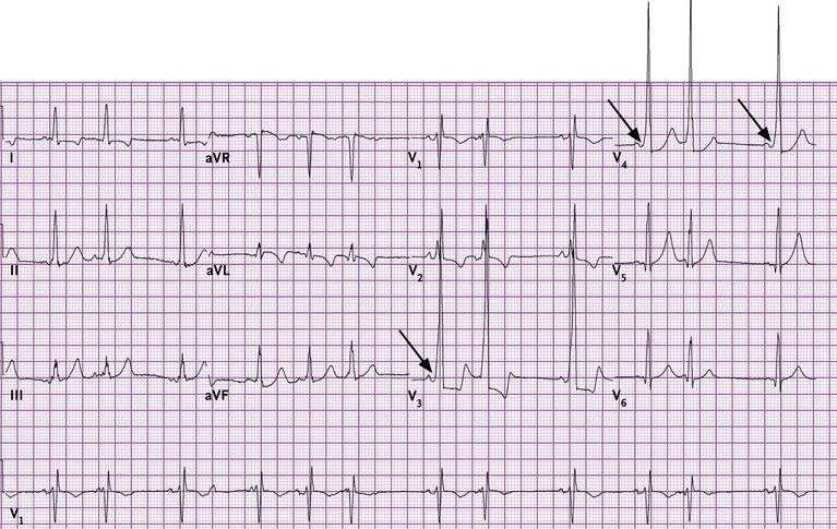
💡 Exam tip: Always check if the short PR is accompanied by a delta wave → this immediately points to WPW syndrome.
| The content on this website is provided for educational and informational purposes only to support exam preparation (e.g., MLA, MRCP, USMLE) and learning. This is NOT medical advice, diagnosis, treatment, or professional guidance. It does not replace consultation with a qualified healthcare professional, official guidelines (e.g., NICE, GMC, BNF), or supervised clinical practice. Always verify information with current, authoritative sources. Makindo and its contributors accept no liability for any reliance on this content, including errors, omissions, or any resulting harm, loss, or consequences. By using this site, you agree to these terms. |
|
|
Categories
- About
- Acute Medicine
- Anaesthetics and Critical Care
- Anatomy
- Anatomy and Physiology
- Biochemistry
- Book
- Cardiology
- Collections
- CompSci
- Crib Sheets
- Critical care
- Dental
- Dermatology
- Differentials
- Drugs
- ENT
- Electrocardiogram
- Embryology
- Emergency Medicine
- Endocrinology
- Ethics
- Foundation Doctors
- GCSE
- Gastroenterology
- General Practice
- Genetics
- Geriatric Medicine
- Geriatrics
- Guidelines
- Haematology
- Hepatology
- Immunology
- Infectious Diseases
- Infographic
- Investigations
- Lists
- MRCP
- Mandatory Training
- Medical Students
- Microbiology
- Nephrology
- Neurology
- Neurosurgery
- Nutrition
- OSCE
- Obstetrics Gynaecology
- Oncology
- Ophthalmology
- Oral Medicine and Dentistry
- Orthopaedics
- Paediatrics
- Palliative
- Palliative Care
- Pathology
- Pharmacology
- Physiology
- Procedures
- Psychiatry
- Public Health
- Radiology
- Respiratory
- Resuscitation
- Revision Topics
- Rheumatology
- Statistics and Research
- Stroke
- Surgery
- Toxicology
- Trauma and Orthopaedics
- USMLE
- Urology
- Vascular Surgery