Makindo Medical Notes"One small step for man, one large step for Makindo" |
|
|---|---|
| Download all this content in the Apps now Android App and Apple iPhone/Pad App | |
| MEDICAL DISCLAIMER: The contents are under continuing development and improvements and despite all efforts may contain errors of omission or fact. This is not to be used for the assessment, diagnosis, or management of patients. It should not be regarded as medical advice by healthcare workers or laypeople. It is for educational purposes only. Please adhere to your local protocols. Use the BNF for drug information. If you are unwell please seek urgent healthcare advice. If you do not accept this then please do not use the website. Makindo Ltd. |
Anatomy of the Hand
-
| About | Anaesthetics and Critical Care | Anatomy | Biochemistry | Cardiology | Clinical Cases | CompSci | Crib | Dermatology | Differentials | Drugs | ENT | Electrocardiogram | Embryology | Emergency Medicine | Endocrinology | Ethics | Foundation Doctors | Gastroenterology | General Information | General Practice | Genetics | Geriatric Medicine | Guidelines | Haematology | Hepatology | Immunology | Infectious Diseases | Infographic | Investigations | Lists | Microbiology | Miscellaneous | Nephrology | Neuroanatomy | Neurology | Nutrition | OSCE | Obstetrics Gynaecology | Oncology | Ophthalmology | Oral Medicine and Dentistry | Paediatrics | Palliative | Pathology | Pharmacology | Physiology | Procedures | Psychiatry | Radiology | Respiratory | Resuscitation | Rheumatology | Statistics and Research | Stroke | Surgery | Toxicology | Trauma and Orthopaedics | Twitter | Urology
Related Subjects: |Anatomy of Skin |Anatomy of the Hand |Anatomy of the Thorax |Anatomy of Muscle Groups |Anatomy of Anatomy of Arteries |Anatomy of Spinal Column
The nerve supply to the hand is primarily provided by three major nerves: the median nerve, the ulnar nerve, and the radial nerve. Each of these nerves is responsible for both sensory and motor innervation, enabling the hand to perform a wide range of functions, from fine motor skills to powerful grips.
Anatomy of the Hand: Bones, Muscles, Functions, and Nerve Supply
- The human hand is a complex and highly functional structure composed of bones, muscles, tendons, ligaments, and nerves. It allows for a wide range of movements and dexterity, enabling tasks from powerful grip to delicate manipulation.
- Understanding the anatomy of the hand is essential for diagnosing and treating injuries and conditions affecting hand function.
Hand movements
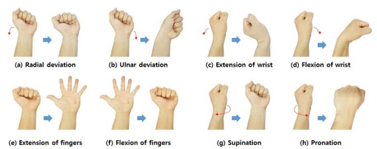
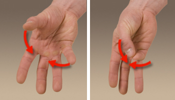 Opposition of the thumb and digits is key for using tools.
Central hand control
- The motor homunculus is a distorted human figure mapped onto the precentral gyrus of the brain, which is the primary motor cortex. It illustrates that body parts requiring precise, fine motor control, such as the hands, face, and tongue, occupy larger areas of the cortex.
- The hand, particularly the fingers, occupies a substantial portion of the motor cortex. This large representation reflects the complex and dexterous movements the hand can perform. Fine motor skills, such as writing, typing, playing musical instruments, and manipulating small objects, demand significant neural control and coordination.
- The density of motor neurons and the richness of the neural connections in this area support rapid and precise adjustments in hand movements.
- The large representation of the hand in the motor cortex is thought to be an evolutionary adaptation that enabled humans to use tools, create art, and perform other activities that require fine motor skills.
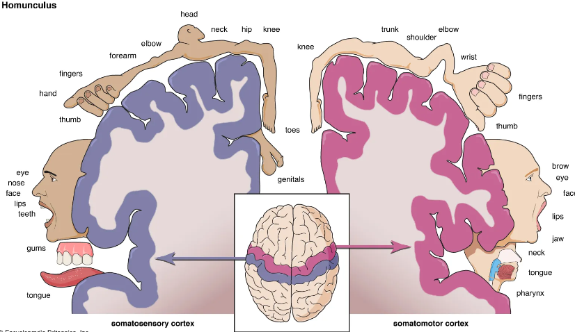
Brain to hand
The corticospinal tract is the primary pathway for transmitting motor commands from the brain to the hand. It involves a sequence of upper motor neurons descending from the motor cortex through the brainstem and spinal cord , where they synapse with lower motor neurons that extend through the brachial plexus and peripheral nerves to the muscles of the hand.
Bones of the Hand
The bones of the hand are divided into three main groups: carpals, metacarpals, and phalanges.
- Carpal Bones (Wrist Bones): Eight small bones arranged in two rows forming the wrist joint.
- Proximal Row (lateral to medial):
- Scaphoid: Boat-shaped; articulates with the radius.
- Lunate: Moon-shaped; articulates with the radius.
- Triquetrum: Pyramidal shape; articulates with the articular disk of the ulna.
- Pisiform: Pea-shaped; sits on top of the triquetrum.
- Distal Row (lateral to medial):
- Trapezium: Articulates with the first metacarpal (thumb).
- Trapezoid: Wedge-shaped; articulates with the second metacarpal.
- Capitate: Largest carpal bone; articulates with the third metacarpal.
- Hamate: Has a hook-like projection (hook of hamate); articulates with the fourth and fifth metacarpals.
- Proximal Row (lateral to medial):
- Metacarpal Bones: Five long bones forming the framework of the palm.
- Numbered I to V from the thumb (lateral) to the little finger (medial).
- Each metacarpal has a base (proximal), shaft, and head (distal).
- Phalanges (Finger Bones): Fourteen bones forming the fingers and thumb.
- Each finger has three phalanges: proximal, middle, and distal.
- The thumb (pollex) has two phalanges: proximal and distal.
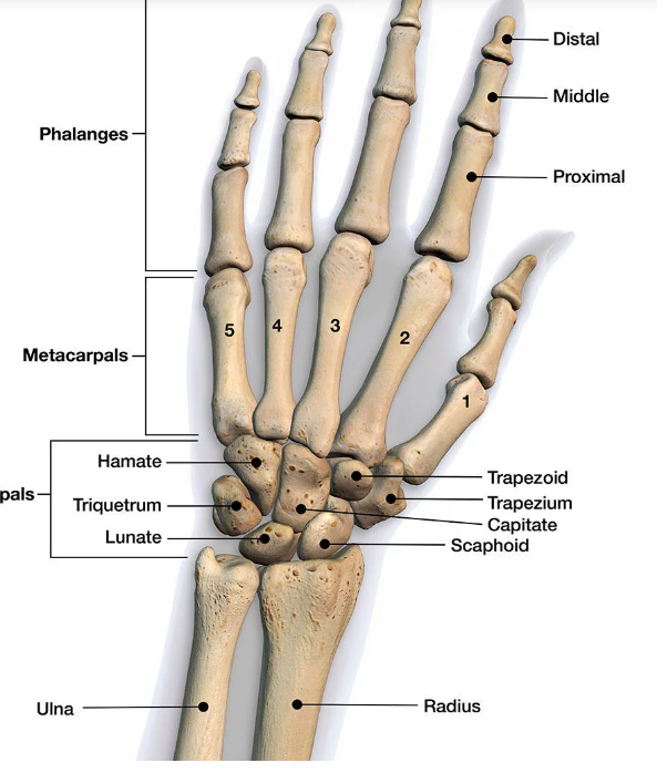
Functions of the Bones
- Support and Structure: Provide the framework for the hand's shape and form.
- Articulation: Form joints that allow for a wide range of movements, including flexion, extension, abduction, adduction, and opposition.
- Attachment Points: Serve as sites for muscle and ligament attachments, facilitating movement and stability.
Muscles of the Hand
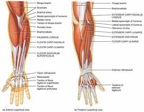
The muscles of the hand are categorized into two groups: extrinsic muscles (originating in the forearm) and intrinsic muscles (originating within the hand). They work together to produce precise and coordinated movements.
Extrinsic Muscles
These muscles originate in the forearm and insert into the hand, controlling gross movements and forceful grips.
- Flexor Muscles (Anterior Forearm):
- Flexor Digitorum Superficialis: Flexes the proximal interphalangeal joints (PIP) of fingers 2–5.
- Flexor Digitorum Profundus: Flexes the distal interphalangeal joints (DIP) of fingers 2–5.
- Flexor Pollicis Longus: Flexes the thumb at the interphalangeal joint.
- Extensor Muscles (Posterior Forearm):
- Extensor Digitorum: Extends fingers 2–5 at the metacarpophalangeal (MCP) and interphalangeal joints.
- Extensor Indicis: Extends the index finger.
- Extensor Digiti Minimi: Extends the little finger.
- Extensor Pollicis Longus and Brevis: Extend the thumb at the MCP and interphalangeal joints.
- Abductor Pollicis Longus: Abducts the thumb at the carpometacarpal joint.
Intrinsic Muscles
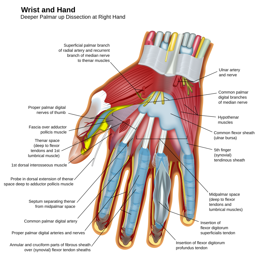
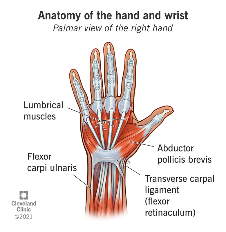
These muscles originate and insert within the hand, allowing fine motor movements and precise control.
- Thenar Muscles (Thumb Side): Form the thenar eminence.
- Abductor Pollicis Brevis: Abducts the thumb.
- Flexor Pollicis Brevis: Flexes the thumb at the MCP joint.
- Opponens Pollicis: Opposes the thumb, allowing it to touch other fingertips.
- Hypothenar Muscles (Little Finger Side): Form the hypothenar eminence.
- Abductor Digiti Minimi: Abducts the little finger.
- Flexor Digiti Minimi Brevis: Flexes the little finger at the MCP joint.
- Opponens Digiti Minimi: Opposes the little finger towards the thumb.
- Lumbricals:
- Four small muscles originating from the tendons of flexor digitorum profundus.
- Flex the MCP joints and extend the PIP and DIP joints of fingers 2–5.
- Interossei Muscles:
- Dorsal Interossei (4 muscles): Abduct fingers 2–4 away from the midline of the hand (middle finger).
- Palmar Interossei (3 muscles): Adduct fingers 2, 4, and 5 towards the midline of the hand.
- Adductor Pollicis:
- Adducts the thumb towards the palm.
Functions of the Muscles
- Gross Movements: Flexion and extension of the wrist and fingers, gripping actions.
- Fine Motor Skills: Precise movements such as typing, writing, and manipulating small objects.
- Opposition: Ability to touch the thumb to other fingertips, crucial for grasping and pinching.
- Abduction and Adduction: Spreading fingers apart and bringing them together.
Nerve Supply of the Hand
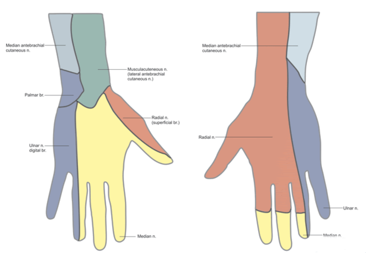
The hand's sensory and motor functions are supplied by three main nerves: the median, ulnar, and radial nerves.
Median Nerve
- Motor Functions:
- Innervates most of the thenar muscles: abductor pollicis brevis, flexor pollicis brevis (superficial head), and opponens pollicis.
- Innervates the lateral two lumbricals (index and middle fingers).
- Sensory Functions:
- Provides sensation to the palmar side of the thumb, index finger, middle finger, and lateral half of the ring finger.
- Also supplies the dorsal tips of the same fingers.
- Clinical Relevance:
- Carpal Tunnel Syndrome: Compression of the median nerve at the wrist leading to numbness, tingling, and muscle weakness.
Ulnar Nerve
- Motor Functions:
- Innervates most of the intrinsic muscles of the hand, including:
- Hypothenar muscles.
- Medial two lumbricals (ring and little fingers).
- All interossei muscles.
- Adductor pollicis.
- Flexor pollicis brevis (deep head).
- Innervates most of the intrinsic muscles of the hand, including:
- Sensory Functions:
- Provides sensation to the palmar and dorsal sides of the little finger and medial half of the ring finger.
- Clinical Relevance:
- Ulnar Nerve Entrapment: Compression at the elbow (cubital tunnel) or wrist (Guyon's canal) causing sensory loss and muscle weakness.
Radial Nerve
- Motor Functions:
- Innervates the extrinsic extensor muscles of the wrist and fingers (posterior compartment of the forearm).
- Does not innervate intrinsic hand muscles.
- Sensory Functions:
- Provides sensation to the dorsal lateral aspect of the hand, including the thumb and proximal portions of the index and middle fingers.
- Clinical Relevance:
- Wrist Drop: Injury to the radial nerve leading to inability to extend the wrist and fingers.
Summary of Nerve Distribution
- Median Nerve: "LOAF" muscles—Lateral two lumbricals, Opponens pollicis, Abductor pollicis brevis, Flexor pollicis brevis (superficial head).
- Ulnar Nerve: All intrinsic muscles not innervated by the median nerve.
- Radial Nerve: All extensor muscles of the wrist and fingers; no intrinsic hand muscles.
Nerves of the Hand and Their Muscle Supply
The following table summarizes the nerves of the hand and the muscles they innervate. This information is essential for understanding the motor functions of the hand and for clinical assessments of nerve injuries.
| Nerve | Muscles Supplied |
|---|---|
| Median Nerve |
|
| Ulnar Nerve |
|
| Radial Nerve |
Note: The radial nerve does not innervate intrinsic hand muscles. |
Summary of Nerve Distribution
- Median Nerve: Supplies most of the thenar muscles and the lateral two lumbricals.
- Ulnar Nerve: Supplies the hypothenar muscles, medial two lumbricals, all interossei muscles, adductor pollicis, deep head of flexor pollicis brevis, and palmaris brevis.
- Radial Nerve: Supplies the extrinsic extensor muscles of the wrist and fingers; does not innervate intrinsic hand muscles.
Additional Details
Median Nerve Motor Innervation:
- Thenar Muscles: Essential for thumb opposition and fine movements.
- Lateral Two Lumbricals: Flex the metacarpophalangeal (MCP) joints and extend the interphalangeal (IP) joints of the index and middle fingers.
Ulnar Nerve Motor Innervation:
- Hypothenar Muscles: Control movements of the little finger.
- Medial Two Lumbricals: Flex the MCP joints and extend the IP joints of the ring and little fingers.
- Interossei Muscles:
- Dorsal Interossei: Abduct fingers 2–4 away from the middle finger.
- Palmar Interossei: Adduct fingers 2, 4, and 5 towards the middle finger.
- Other Muscles:
- Adductor Pollicis: Adducts the thumb towards the palm.
- Flexor Pollicis Brevis (deep head): Assists in flexing the thumb.
- Palmaris Brevis: Wrinkles the skin of the hypothenar eminence and deepens the hollow of the palm.
Radial Nerve Motor Innervation:
- Controls extension of the wrist and fingers, enabling opening of the hand and release of objects.
- Innervates muscles that extend and abduct the thumb.
- Clinical Note: Injury to the radial nerve can lead to "wrist drop," where the patient cannot extend the wrist and fingers.
Conclusion
The intricate anatomy of the hand's bones, muscles, and nerves allows for its remarkable functionality. The coordination between the skeletal structure and muscular movements, combined with precise nerve control, enables the hand to perform complex tasks essential for daily activities. Understanding this anatomy is vital for healthcare professionals in diagnosing injuries, managing conditions, and performing surgical interventions.