Makindo Medical Notes"One small step for man, one large step for Makindo" |
|
|---|---|
| Download all this content in the Apps now Android App and Apple iPhone/Pad App | |
| MEDICAL DISCLAIMER: The contents are under continuing development and improvements and despite all efforts may contain errors of omission or fact. This is not to be used for the assessment, diagnosis, or management of patients. It should not be regarded as medical advice by healthcare workers or laypeople. It is for educational purposes only. Please adhere to your local protocols. Use the BNF for drug information. If you are unwell please seek urgent healthcare advice. If you do not accept this then please do not use the website. Makindo Ltd. |
Stroke and Vision
-
| About | Anaesthetics and Critical Care | Anatomy | Biochemistry | Cardiology | Clinical Cases | CompSci | Crib | Dermatology | Differentials | Drugs | ENT | Electrocardiogram | Embryology | Emergency Medicine | Endocrinology | Ethics | Foundation Doctors | Gastroenterology | General Information | General Practice | Genetics | Geriatric Medicine | Guidelines | Haematology | Hepatology | Immunology | Infectious Diseases | Infographic | Investigations | Lists | Microbiology | Miscellaneous | Nephrology | Neuroanatomy | Neurology | Nutrition | OSCE | Obstetrics Gynaecology | Oncology | Ophthalmology | Oral Medicine and Dentistry | Paediatrics | Palliative | Pathology | Pharmacology | Physiology | Procedures | Psychiatry | Radiology | Respiratory | Resuscitation | Rheumatology | Statistics and Research | Stroke | Surgery | Toxicology | Trauma and Orthopaedics | Twitter | Urology
Related Subjects: |Anatomy the Medulla Oblongata |Anatomy of the Midbrain |Anatomy of the Pons
🧠 Introduction
🔹 About 30% of stroke patients develop post-stroke visual impairment. 🔹 Common symptoms: hemianopia, neglect, diplopia, ↓ visual acuity, ptosis, anisocoria, nystagmus. 🔹 Homonymous hemianopia affects ~8% of stroke patients – some may still be driving.
🧬 Aetiology
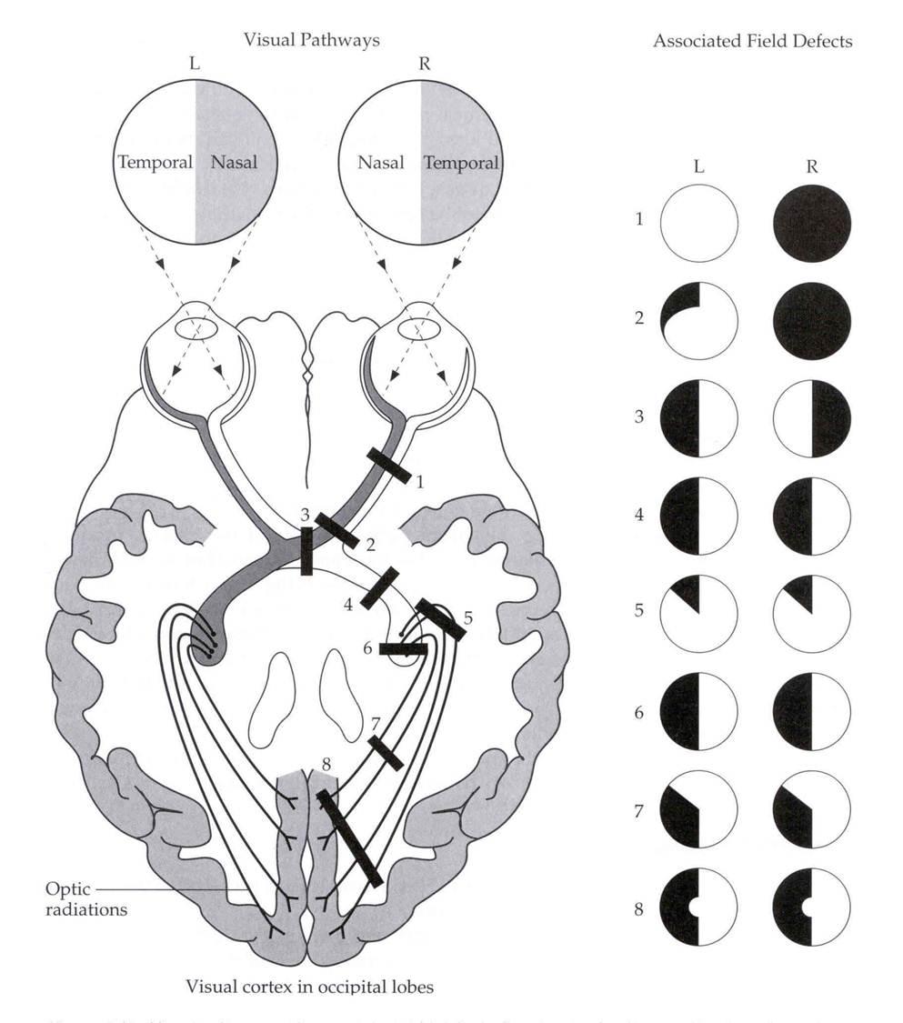
- 👁️ Homonymous hemianopia: Loss of the same right/left halves of the visual field in both eyes. ➡️ Cause: Stroke of optic radiation or occipital cortex (MCA or PCA infarcts). ➡️ Macular sparing may occur if dual MCA/PCA supply to occipital pole.
- ◼️ Quadrantanopia:
- 🌙 Superior quadrantanopia: Temporal lobe lesion (“pie in the sky”).
- 🌄 Inferior quadrantanopia: Parietal lobe lesion (“pie on the floor”).

📊 Range of Visual Field Losses
| Visual Loss | Cause & Summary |
|---|---|
| Altitudinal defect | BRAO, optic neuropathy, retinal detachment, glaucoma |
| Central scotoma | Macular disease, optic neuritis/atrophy |
| Monocular blindness | CRAO, GCA, PMR, or vascular risk factors |
| Monocular quadrantanopia | Branch retinal artery occlusion |
| Dynamic loss (scotoma, aura) | Migraine with aura |
| Bitemporal hemianopia | Pituitary tumour, craniopharyngioma → MRI required |
| Lower homonymous quadrantanopia | Contralateral temporal lobe stroke |
| Upper homonymous quadrantanopia | Contralateral parietal lobe stroke |
| Complete homonymous hemianopia + motor/sensory deficit | Large MCA infarct |
| Complete homonymous hemianopia (no motor deficit) | PCA occipital infarct |
| Complete blindness | Rare, unless multiple bilateral lesions |
| Prosopagnosia | Inability to recognize faces (bilateral inferior occipital/temporal lesions) |
👁️ Left Homonymous Hemianopia (Patient’s View)

🔍 Visual Neglect
🧭 A parietal lobe syndrome → patients ignore one side (usually left). 🪒 May shave/eat only one side. 💡 Managed with occupational therapy & compensatory scanning strategies.
📖 Hemianopic Alexia
📚 Reading disorder post-stroke, usually left hemisphere → disrupts right visual field used for guiding eye movements. 🚨 Common, distressing, requires rehab input.
🚗 Driving & Visual Field Defects
Complete hemianopia = unsafe to drive. DVLA guidelines require assessment before resuming driving. DVLA guidance.
📏 DVLA Minimum Standards for Group 1 Drivers
- 120° horizontal field (≥50° left & right).
- No binocular defect within 20° of fixation.
- Homonymous/bitemporal defects close to fixation = not permitted.
DVLA testing includes: ✔️ Binocular Esterman field test (standard) ✔️ Monocular full-field charts (specific cases) ✔️ Goldmann perimetry (rare, strict criteria)
💊 Management of Visual Field Defects
- Multidisciplinary rehab: ophthalmology, orthoptics, OT, stroke physicians.
- Education + compensatory scanning strategies.
- Prism lenses → shift visual field.
- Refer to patient resources (Chest Heart & Stroke Scotland sheet).
- Prognosis variable → reassess at 2–3 months.
📚 References
- [1] Gilhotra JS, Mitchell P, Healey PR, et al. Homonymous visual field defects and stroke. Stroke 2002;33:2417-20.
- [2] Visual field defects after stroke. Aust Fam Physician, 2010.
- [3] DVLA Standards for Fitness to Drive.