
| Download the amazing global Makindo app: ✅ Means NICE/National Guidelines 2026 compliant Android | Apple | |
|---|---|
| MEDICAL DISCLAIMER: Educational use only. Not for diagnosis or management. See below for full disclaimer. |
Dermatomyositis ✅
Related Subjects: |Relapsing Polychondritis |Reactive Arthritis |Raynaud's Phenomenon |Polymyositis |Dermatomyositis |Polyarteritis nodosa |Osteoporosis |Rheumatoid Arthritis |Systemic Sclerosis (Scleroderma) |Rheumatology Autoantibodies |Overlap Syndrome |Inclusion Body Myositis |Inflammatory Myopathies |Psoriatic Arthritis |Adult Onset Still's Disease |Alkaptonuria |Behcet's Syndrome
🌸 Dermatomyositis is an idiopathic inflammatory myopathy with characteristic cutaneous features and variable skeletal muscle involvement. Typical features include symmetrical proximal muscle weakness, a heliotrope rash (violaceous periorbital rash), and Gottron’s papules/sign over the MCP/PIP joints and extensor surfaces. ⚠️ In adult-onset disease, always think about associated malignancy, especially in higher-risk phenotypes.
🧬 About
- Immune-mediated inflammatory myopathy with prominent microangiopathy / vasculopathy, skin disease, and possible systemic involvement.
- May affect muscle, skin, lungs, swallowing, and occasionally the heart.
- Adult-onset dermatomyositis has a recognised association with occult malignancy; risk is highest around the time of disease onset.
📊 Epidemiology
- Rare condition; more common in women.
- Occurs in both children and adults.
- Adult-onset disease carries malignancy risk; juvenile dermatomyositis is not routinely associated with cancer.
🎗️ Malignancy Association
- Think particularly about malignancy in adult-onset disease, especially if there is older age at onset, male sex, dysphagia, cutaneous necrosis/ulceration, rapid onset, poor response to immunosuppression, or anti-TIF1-γ / anti-NXP2 positivity.
- Common associated cancers include lung, ovarian, breast, colorectal/gastrointestinal, and lymphoma.
- Nasopharyngeal carcinoma is particularly relevant in some East and South-East Asian populations.
- Continue routine age- and sex-appropriate national cancer screening as well as targeted assessment where indicated.
🩺 Clinical Features
- 💪 Symmetrical proximal muscle weakness – difficulty climbing stairs, rising from a chair, washing hair, or lifting arms overhead.
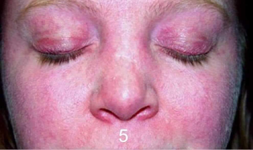
- 🌸 Skin signs:
- Heliotrope rash with periorbital oedema.
- Gottron’s papules/sign over knuckles, elbows, or knees.
- Shawl sign / V-sign – photosensitive erythema over shoulders, upper chest, or back.
- Mechanic’s hands – hyperkeratotic, cracked fingertips (especially with antisynthetase overlap).
- Nailfold telangiectasia.
- Scalp erythema/pruritus may occur.
- 🫁 Lung involvement: interstitial lung disease, especially in antisynthetase or anti-MDA5 phenotypes.
- 🍽️ Dysphagia may occur and is clinically important because of weight loss and aspiration risk.
- ❤️ Occasionally associated with cardiac involvement.
- ⚖️ Constitutional features may include fatigue, weight loss, arthralgia, and reduced exercise tolerance.
🔬 Investigations
- Bloods: CK may be raised (but can be normal, especially in some phenotypes); also check AST/ALT, LDH, CRP/ESR, FBC, U&Es, LFTs, bone profile.
- Autoantibodies: request a myositis-specific / myositis-associated antibody panel.
- Anti-TIF1-γ, anti-NXP2 → malignancy risk markers in adult-onset disease.
- Anti-MDA5 → risk of rapidly progressive ILD.
- Anti-Mi-2 → classic cutaneous dermatomyositis phenotype.
- Anti-Jo-1 and other antisynthetase antibodies → antisynthetase syndrome / ILD / mechanic’s hands / Raynaud’s.
- MRI muscle: useful to identify inflamed muscle and guide biopsy.
- EMG: supports a myopathic process but is not diagnostic on its own.
- Muscle biopsy: classically shows perifascicular atrophy with perivascular/perimysial inflammation.
- Skin biopsy: may support the diagnosis where rash is prominent.
- Screen for ILD in higher-risk patients with chest imaging, pulmonary function tests (including gas transfer), and HRCT where indicated.
- Assess swallowing if dysphagia is suspected.
- Consider cardiac screening with ECG, echocardiography, and troponin I if clinically indicated.
- Malignancy screen: tailored to risk profile; in higher-risk adult patients consider CT thorax/abdomen/pelvis and further targeted tests as indicated.
💊 Management
- 🤝 Specialist MDT care – usually rheumatology ± neurology, dermatology, respiratory, SALT, physiotherapy, and oncology where needed.
- 📉 Glucocorticoids are usually first-line induction therapy.
- 🛡️ Add a steroid-sparing immunosuppressant early in many patients (for example methotrexate, azathioprine, mycophenolate, tacrolimus/ciclosporin depending on phenotype and organ involvement).
- 💉 Refractory / severe disease: consider IV methylprednisolone, IVIG, rituximab, or cyclophosphamide in specialist care.
- 🌞 Sun protection is important for cutaneous disease.
- 🏃 Exercise / rehabilitation with specialist physiotherapy or OT should be part of routine care.
- 🦴 Assess bone protection if prolonged steroids are used.
- 🍽️ Actively manage dysphagia, aspiration risk, nutrition, and respiratory complications.
🧾 Comparison of Inflammatory Myopathies
| Feature | 💉 Polymyositis* | 🌸 Dermatomyositis | 🧓 Inclusion Body Myositis |
|---|---|---|---|
| Typical age | Adults | Children or adults | > 50 years |
| Weakness pattern | Proximal | Proximal ± skin disease | Finger flexors + quadriceps, often asymmetric |
| Skin changes | No | Yes – heliotrope rash, Gottron’s papules/sign | No |
| CK | Usually raised | Usually raised, but may be normal | Normal or mildly raised |
| Cancer association | Less clear / lower than DM | Important association in adult-onset disease | Not a prominent classic association |
| Biopsy | Endomysial inflammation with CD8+ T cells | Perifascicular atrophy; perivascular/perimysial inflammation | Endomysial inflammation + rimmed vacuoles |
| Response to treatment | Variable | Often responsive to immunosuppression | Poor |
Teaching point 🩺: In adult-onset dermatomyositis, think in three parallel directions: muscle disease, skin disease, and systemic associations (especially malignancy, ILD, and dysphagia). Anti-Jo-1 is more suggestive of an antisynthetase phenotype than “classic dermatomyositis”, while anti-TIF1-γ and anti-NXP2 are more helpful when thinking about cancer risk.
*Pure polymyositis is now considered much less common than older textbooks suggested, and modern practice often reclassifies patients into other inflammatory myopathy subtypes.
:100%;">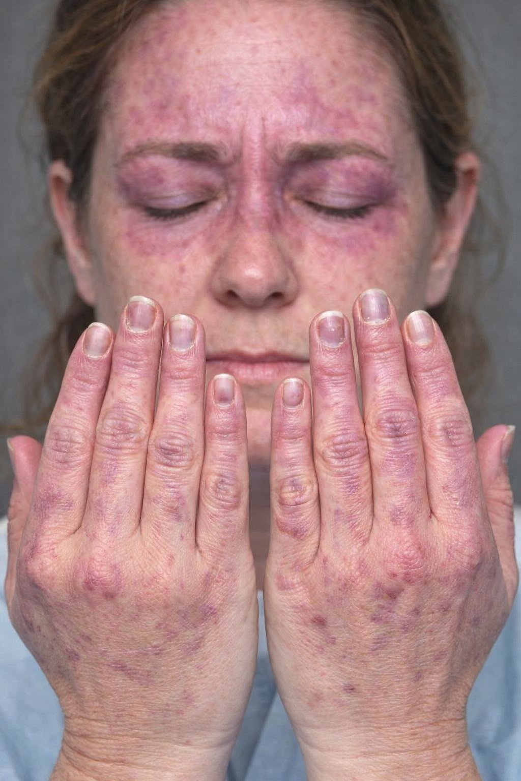
| The content on this website is provided for educational and informational purposes only to support exam preparation (e.g., MLA, MRCP, USMLE) and learning. This is NOT medical advice, diagnosis, treatment, or professional guidance. It does not replace consultation with a qualified healthcare professional, official guidelines (e.g., NICE, GMC, BNF), or supervised clinical practice. Always verify information with current, authoritative sources. Makindo and its contributors accept no liability for any reliance on this content, including errors, omissions, or any resulting harm, loss, or consequences. By using this site, you agree to these terms. |
|
|
Categories
- About
- Acute Medicine
- Anaesthetics and Critical Care
- Anatomy
- Anatomy and Physiology
- Biochemistry
- Book
- Cardiology
- Collections
- CompSci
- Crib Sheets
- Critical care
- Dental
- Dermatology
- Differentials
- Drugs
- ENT
- Electrocardiogram
- Embryology
- Emergency Medicine
- Endocrinology
- Ethics
- Foundation Doctors
- GCSE
- Gastroenterology
- General Practice
- Genetics
- Geriatric Medicine
- Geriatrics
- Guidelines
- Haematology
- Hepatology
- Immunology
- Infectious Diseases
- Infographic
- Investigations
- Lists
- MRCP
- Mandatory Training
- Medical Students
- Microbiology
- Nephrology
- Neurology
- Neurosurgery
- Nutrition
- OSCE
- Obstetrics Gynaecology
- Oncology
- Ophthalmology
- Oral Medicine and Dentistry
- Orthopaedics
- Paediatrics
- Palliative
- Palliative Care
- Pathology
- Pharmacology
- Physiology
- Procedures
- Psychiatry
- Public Health
- Radiology
- Respiratory
- Resuscitation
- Revision Topics
- Rheumatology
- Statistics and Research
- Stroke
- Surgery
- Toxicology
- Trauma and Orthopaedics
- USMLE
- Urology
- Vascular Surgery