
| Download the amazing global Makindo app: ✅ Means NICE/National Guidelines 2026 compliant Android | Apple | |
|---|---|
| MEDICAL DISCLAIMER: Educational use only. Not for diagnosis or management. See below for full disclaimer. |
Necrotising fasciitis
Related Subjects: |Cellulitis |Pyoderma gangrenosum |Pemphigus Vulgaris |Toxic Epidermal Necrolysis |Stevens-Johnson Syndrome |Necrotising fasciitis |Gas Gangrene (Clostridium perfringens) |Purpura Fulminans |Severe burns |Anatomy of Skin
💡 Teaching Pearl: Necrotising fasciitis is both a medical and surgical emergency. Immediate diagnosis and treatment save lives. Delay in surgery = higher mortality. Variants: Meleney’s gangrene, Fournier’s gangrene.
| ⚠️ Necrotising Fasciitis – Key Emergency Features |
|---|
|
Necrotizing fasciitis is a medical and surgical emergency that requires immediate attention. Prompt diagnosis and treatment are crucial, as delays can significantly increase mortality rates. This severe infection often necessitates extensive surgery, reconstruction, and, in some cases, amputation. Meleney’s synergistic gangrene and Fournier’s gangrene are variants of a similar disease process.
ℹ️ About
- Definition: Necrotizing fasciitis is a deep infection characterized by necrosis and damage to the dermis and subcutaneous tissue.
- Pathophysiology: The infection can traverse the usual fascial layers that typically limit the spread of infection.
- Etiology: Caused by Gram-negative or Gram-positive anaerobic bacteria.
- Entry Point: May originate from a small wound at the skin surface.
Risks
- Individuals with diabetes mellitus.
- People who inject drugs.
- Patients with haematological malignancies.
- Smoking; penetrating trauma
- Pressure sores; immunosuppression
- Perineal infection (perianal abscess, Bartholin’s cysts)
- Skin damage/infection (abrasions, bites, boils)
Microbiology: Polymicrobial and Mixed
Necrotising fasciitis results from synergistic, polymicrobial infection; most commonly a streptococcal species (Group A -haemolytic) in combination with Staphylococcus, Escherichia coli, Pseudomonas, Proteus, Bacteroides or Clostridia
- Group A Beta-Haemolytic Streptococci (GAS): A major cause, often referred to as "flesh-eating disease."
- Staphylococcus aureus (including MRSA): Commonly involved in mixed infections.
- Aeromonas hydrophila: Predominantly seen in tropical regions.
- Vibrio vulnificus: Associated with seawater exposure in tropical climates.
- Fungal Infections: Can occur, especially in immunocompromised individuals.
- Clostridium Species: Associated with gas gangrene and gas formation in tissues.
Classification
- Type 1: Mixed Infection
- Combination of Enterobacteriaceae and anaerobes.
- Common in immunocompromised patients, diabetics, and post-surgical patients.
- Type 2: Streptococcal Infection
- Caused by Streptococcus pyogenes (Lancefield Group A).
- Produces a superantigen that activates T cells and macrophages, leading to extensive tissue damage.
- Known as the "flesh-eating disease."
- Type 3: Clostridial Myonecrosis (Gas Gangrene)
- Related to trauma or surgery.
- Caused by Clostridium perfringens.
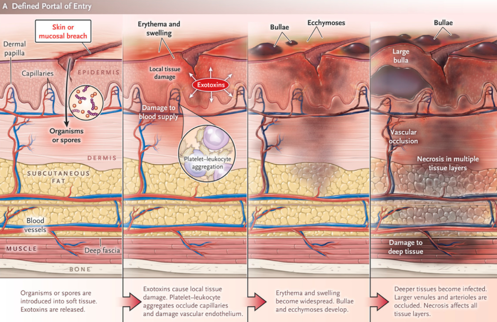
🩺 Clinical Features
Oedema stretching beyond visible skin erythema; a woody-hard texture to the subcutaneous tissues; an inability to distinguish fascial planes and muscle groups on palpation; disproportionate pain in relation to the affected area, with associated skin vesicles and soft tissue crepitus
- Severe Pain and Swelling: Intense pain and rapid swelling in the affected area.
- Systemic Symptoms: Severe systemic inflammatory response, including fever and tachycardia.
- Anaesthetic Center: The center of the lesion may be anaesthetic due to nerve damage.
- Crepitus: Presence of gas beneath the skin, detectable as crepitus.
- Multisystem Failure: Advanced cases can lead to multi-organ dysfunction and failure.
Laboratory Risk Indicator for Necrotizing Fasciitis (LRINEC) Score
- C-Reactive Protein (CRP): >150 mg/L or 15 mg/dL (+4)
- White Cell Count (WCC): 15-25 (+1), >25 (+2)
- Sodium (Na): <135 mmol/L (+2)
- Creatinine: >141 μmol/L or 1.6 mg/dL (+2)
- Glucose: >180 mg/dL or 100 mmol/L (+1)
- Interpretation:
- A LRINEC score of 6 or more suggests necrotizing fasciitis.
- A LRINEC score below 6 does not rule out the diagnosis.
- Note: Use with caution as the LRINEC Score has performed poorly in external validation. MDCalc - LRINEC Score
Differential Diagnosis
- Toxic Shock Syndrome.
- Mucormycosis Infection.
🔎 Investigations
- Complete Blood Count (CBC): Elevated WCC.
- C-Reactive Protein (CRP): Significantly elevated.
- Urea and Electrolytes (U&E): May reveal acute kidney injury (AKI).
- Calcium Levels: Hypocalcemia may be present.
- Imaging: MRI or CT scans can assess the extent of soft tissue involvement and presence of gas.
💊 Management
🚨 Necrotising fasciitis is a surgical emergency. If the diagnosis is suspected, obtain immediate senior surgical review. Do not delay operative exploration and debridement while waiting for CT/MRI, microbiology results or a LRINEC score. Early and complete surgical source control is the most important determinant of survival.
- 🚑 Immediate Resuscitation – ABCDE
- Assess and stabilise Airway, Breathing and Circulation. Give oxygen if hypoxaemic and support ventilation where required. Obtain large-bore IV access and begin IV crystalloid resuscitation if hypotensive or hypoperfused.
- Measure lactate and manage as sepsis/septic shock where appropriate. If hypotension persists despite adequate fluid resuscitation, commence vasopressor support — usually noradrenaline — in critical care.
- Monitor urine output, renal function, electrolytes, glucose, coagulation and other evidence of evolving organ dysfunction. Early involvement of critical care, microbiology/infectious diseases and the appropriate surgical specialty is recommended.
- 🔪 Emergency Surgical Source Control
- Urgent surgical exploration and aggressive debridement are the definitive treatment.
- Remove all necrotic and non-viable skin, subcutaneous tissue, fascia and muscle until healthy bleeding tissue is reached.
- Surgical exploration is both diagnostic and therapeutic; classical findings include grey necrotic fascia, thin “dishwater” fluid, poor tissue resistance and easy blunt dissection along fascial planes.
- Do not delay theatre for imaging when clinical suspicion is high.
- Send multiple deep tissue samples for Gram stain, culture and antimicrobial sensitivities; superficial wound swabs are much less useful.
- Obtain blood cultures where possible, but this must not delay antibiotics or surgery.
- Patients frequently require repeat operative exploration and debridement, commonly within approximately 24 hours and thereafter until no further necrotic tissue remains.
- Occasionally extensive tissue destruction necessitates amputation to achieve adequate source control.
- 💉 Immediate IV Antibiotics
- Give broad-spectrum IV antibiotics immediately once the condition is suspected.
- Initial therapy must cover:
- Group A streptococci and other streptococci
- Staphylococcus aureus
- Gram-negative organisms
- Anaerobes
- MRSA where epidemiological or patient-specific risk warrants it
- Typical UK empirical approach: an anti-pseudomonal broad-spectrum β-lactam such as piperacillin–tazobactam together with clindamycin, with additional MRSA cover when indicated.
- Important: exact antibiotic choice and dosing vary between NHS Trusts — follow the local severe skin/soft-tissue infection guideline and discuss urgently with Microbiology.
- Where Group A streptococcal necrotising fasciitis is confirmed, treatment generally includes a β-lactam such as benzylpenicillin plus clindamycin.
- Why clindamycin? It suppresses bacterial protein synthesis and therefore reduces production of streptococcal and clostridial exotoxins. This effect is valuable even when the bacterial burden is high or organisms are relatively stationary in growth phase.
- Once microbiological results are available, narrow antimicrobial therapy according to the organism and sensitivities.
- 🦠 Streptococcal Toxic Shock Syndrome
- Suspect this when invasive Group A streptococcal infection is associated with shock and multiorgan failure. Urgent source control, β-lactam therapy and a toxin-suppressing agent such as clindamycin are central to treatment.
- IV immunoglobulin (IVIG) may be considered in severe streptococcal toxic shock syndrome after discussion with Critical Care and Microbiology/Infectious Diseases, although evidence of benefit remains uncertain.
- 🩹 Wound Management and Reconstruction
- Following adequate debridement, large open wounds require meticulous wound care. Negative-pressure wound therapy may be useful once infection and necrosis have been adequately controlled.
- Early involvement of plastic and reconstructive surgery may be required. Skin grafts, local flaps or free-tissue transfer may subsequently be needed.
- Early nutritional support is important because extensive wounds and repeated surgery produce major protein and metabolic losses.
- ⚠️ Hyperbaric Oxygen
- Hyperbaric oxygen is not part of routine initial treatment. Evidence for benefit is uncertain and treatment must never delay resuscitation, antibiotics or surgical debridement.
- It may occasionally be considered in specialist centres as an adjunct in selected cases, particularly some clostridial infections, but only after definitive surgical management has been organised.
💡 Clinical Pearl The critical therapeutic sequence is: Recognise → Resuscitate → Antibiotics → Theatre → Debride → Re-look.
A patient with severe pain, systemic toxicity and rapidly progressive soft-tissue inflammation should be treated as having necrotising fasciitis until proven otherwise. A normal scan, low LRINEC score or absence of gas does not safely exclude the diagnosis.
📈 Prognosis
- Necrotising fasciitis carries substantial morbidity and mortality, particularly when associated with shock or delayed source control. Any Delay to surgical debridement is associated with worse outcomes.
- Major complications include septic shock, acute kidney injury, respiratory failure, DIC, multiorgan failure and extensive tissue loss. Survivors may require repeated operations, skin grafting, reconstructive surgery, rehabilitation and occasionally amputation. Functional and psychological consequences can persist long after the acute infection has resolved.
✅ Key Point
Necrotising fasciitis is fundamentally a surgical disease. Antibiotics and intensive-care support are essential, but survival depends on recognising the condition early and achieving rapid, radical surgical source control.
| The content on this website is provided for educational and informational purposes only to support exam preparation (e.g., MLA, MRCP, USMLE) and learning. This is NOT medical advice, diagnosis, treatment, or professional guidance. It does not replace consultation with a qualified healthcare professional, official guidelines (e.g., NICE, GMC, BNF), or supervised clinical practice. Always verify information with current, authoritative sources. Makindo and its contributors accept no liability for any reliance on this content, including errors, omissions, or any resulting harm, loss, or consequences. By using this site, you agree to these terms. |
|
|
Categories
- About
- Acute Medicine
- Anaesthetics and Critical Care
- Anatomy
- Anatomy and Physiology
- Biochemistry
- Book
- Cardiology
- Collections
- CompSci
- Crib Sheets
- Critical care
- Dental
- Dermatology
- Differentials
- Drugs
- ENT
- Electrocardiogram
- Embryology
- Emergency Medicine
- Endocrinology
- Ethics
- Foundation Doctors
- GCSE
- Gastroenterology
- General Practice
- Genetics
- Geriatric Medicine
- Geriatrics
- Guidelines
- Haematology
- Hepatology
- Immunology
- Infectious Diseases
- Infographic
- Investigations
- Lists
- MRCP
- Mandatory Training
- Medical Students
- Microbiology
- Nephrology
- Neurology
- Neurosurgery
- Nutrition
- OSCE
- Obstetrics Gynaecology
- Oncology
- Ophthalmology
- Oral Medicine and Dentistry
- Orthopaedics
- Paediatrics
- Palliative
- Palliative Care
- Pathology
- Pharmacology
- Physiology
- Procedures
- Psychiatry
- Public Health
- Radiology
- Respiratory
- Resuscitation
- Revision Topics
- Rheumatology
- Statistics and Research
- Stroke
- Surgery
- Toxicology
- Trauma and Orthopaedics
- USMLE
- Urology
- Vascular Surgery