Makindo Medical Notes"One small step for man, one large step for Makindo" |

|
|---|---|
| Download all this content in the Apps now Android App and Apple iPhone/Pad App | |
| MEDICAL DISCLAIMER: The contents are under continuing development and improvements and despite all efforts may contain errors of omission or fact. This is not to be used for the assessment, diagnosis, or management of patients. It should not be regarded as medical advice by healthcare workers or laypeople. It is for educational purposes only. Please adhere to your local protocols. Use the BNF for drug information. If you are unwell please seek urgent healthcare advice. If you do not accept this then please do not use the website. Makindo Ltd. | |
Laryngeal anatomy
-
| About | Anaesthetics and Critical Care | Anatomy | Biochemistry | Cardiology | Clinical Cases | CompSci | Crib | Dermatology | Differentials | Drugs | ENT | Electrocardiogram | Embryology | Emergency Medicine | Endocrinology | Ethics | Foundation Doctors | Gastroenterology | General Information | General Practice | Genetics | Geriatric Medicine | Guidelines | Haematology | Hepatology | Immunology | Infectious Diseases | Infographic | Investigations | Lists | Microbiology | Miscellaneous | Nephrology | Neuroanatomy | Neurology | Nutrition | OSCE | Obstetrics Gynaecology | Oncology | Ophthalmology | Oral Medicine and Dentistry | Paediatrics | Palliative | Pathology | Pharmacology | Physiology | Procedures | Psychiatry | Radiology | Respiratory | Resuscitation | Rheumatology | Statistics and Research | Stroke | Surgery | Toxicology | Trauma and Orthopaedics | Twitter | Urology
Anatomy of the Larynx
The larynx, commonly known as the voice box, is a vital structure located in the anterior part of the neck, situated between the pharynx and the trachea. It plays an essential role in breathing, phonation (voice production), and protecting the airway during swallowing. The larynx is composed of several cartilages, muscles, ligaments, and membranes that work together to control airflow and sound production.

Cartilages of the Larynx
The larynx consists of nine cartilages, three of which are single, and three are paired.
- Single Cartilages:
- Thyroid Cartilage: The largest cartilage of the larynx, it forms the Adam's apple and serves as a protective shield for the vocal cords. It has a prominent anterior projection called the laryngeal prominence.
- Cricoid Cartilage: Located below the thyroid cartilage, the cricoid cartilage is shaped like a ring and provides structural support. It forms a complete ring around the airway.
- Epiglottis: A leaf-shaped cartilage that acts as a lid for the larynx, preventing food and liquids from entering the airway during swallowing.
- Paired Cartilages:
- Arytenoid Cartilages: Pyramid-shaped cartilages located at the posterior end of the larynx. They are crucial for vocal cord movement and positioning.
- Corniculate Cartilages: Small horn-shaped cartilages that sit atop the arytenoid cartilages, aiding in vocal cord tension and movement.
- Cuneiform Cartilages: Small, rod-shaped cartilages embedded in the aryepiglottic folds. They provide support to the vocal folds and laryngeal structure.

Muscles of the Larynx
The muscles of the larynx are divided into two main groups: intrinsic and extrinsic muscles.
- Intrinsic Muscles:
- Cricoarytenoid Muscles:
- Lateral cricoarytenoid: Adducts the vocal cords, bringing them together for phonation.
- Posterior cricoarytenoid: The only muscle responsible for abducting the vocal cords, allowing for breathing.
- Thyroarytenoid Muscle: Relaxes the vocal cords, shortening and thickening them, which lowers the pitch of the voice.
- Cricothyroid Muscle: Stretches and tenses the vocal cords, raising the pitch of the voice.
- Transverse and Oblique Arytenoid Muscles: Work to close the posterior part of the glottis, facilitating vocal cord adduction during speech and swallowing.
- Cricoarytenoid Muscles:
- Extrinsic Muscles:
- Suprahyoid Muscles: These muscles elevate the larynx during swallowing. Examples include the digastric, mylohyoid, and stylohyoid muscles.
- Infrahyoid Muscles: Also called "strap muscles," they depress the larynx after swallowing. Examples include the sternohyoid, omohyoid, and sternothyroid muscles.

Vocal Cords (Vocal Folds)
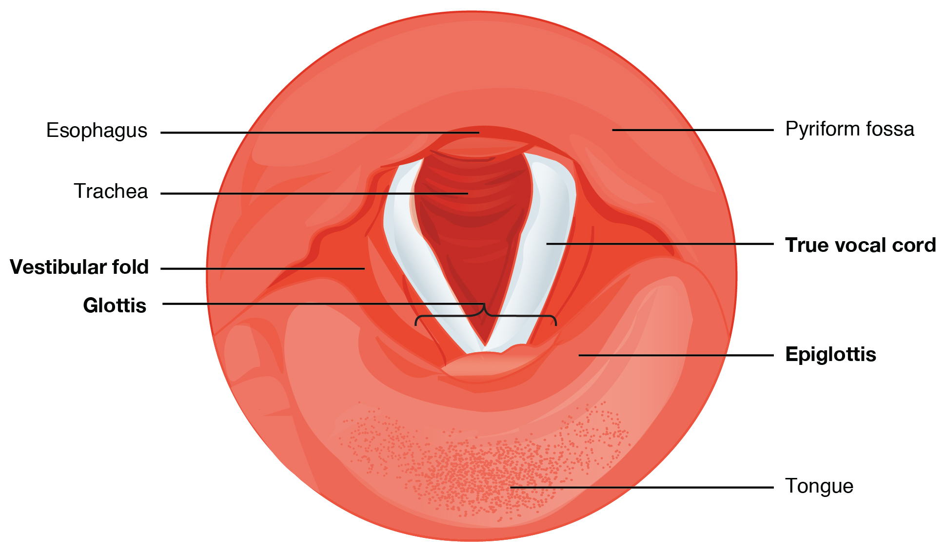
The vocal cords, also known as vocal folds, are twin infoldings of mucous membrane stretched horizontally across the larynx. They are responsible for sound production.
- True Vocal Cords: These are the main vocal folds that produce sound when air passes through them and they vibrate. The tension and position of the cords determine pitch and tone.
- False Vocal Cords (Vestibular Folds): Located above the true vocal cords, they do not contribute to sound production but play a role in protecting the airway and holding breath during activities like lifting heavy objects.
Functions of the Larynx
- Phonation: The primary function of the larynx is to produce sound. This is achieved by the vibration of the vocal cords as air passes through them. Muscles in the larynx control the tension and position of the vocal cords, altering pitch and volume.
- Breathing: The larynx allows the passage of air between the pharynx and trachea. The vocal cords can open widely (abduction) to allow free flow of air during breathing.
- Protection of the Airway: During swallowing, the larynx elevates, and the epiglottis folds down over the glottis (the opening between the vocal cords) to prevent food and liquid from entering the trachea.
- Swallowing: The larynx elevates during swallowing to direct food and liquids into the esophagus, while the epiglottis closes off the airway to prevent aspiration.
- Cough Reflex: The larynx plays a key role in initiating the cough reflex, which helps clear the airway of irritants or blockages.
Blood Supply of the Larynx
- Arteries: The larynx is supplied by the superior laryngeal artery (a branch of the superior thyroid artery) and the inferior laryngeal artery (a branch of the inferior thyroid artery).
- Veins: Venous drainage is through the superior and inferior laryngeal veins, which drain into the superior and inferior thyroid veins.
Nerve Supply of the Larynx
- Motor Innervation:
The muscles of the larynx are innervated by branches of the vagus nerve (CN X):
- Recurrent Laryngeal Nerve: Supplies all intrinsic muscles of the larynx except the cricothyroid muscle.
- Superior Laryngeal Nerve: Supplies the cricothyroid muscle, which is involved in tension and lengthening of the vocal cords.
- Sensory Innervation: The internal branch of the superior laryngeal nerve provides sensory innervation to the mucosa above the vocal cords, while the recurrent laryngeal nerve provides sensory input below the vocal cords.